Anatomy Of The Heart Drawing
Anatomy Of The Heart Drawing - Others are based on an adapted 3d model (texturing and modelling) to the study of anatomy. Draw the main shape of your human heart drawing begin this tutorial, by drawing the main shape of the human heart represented by a tilted triangle. Add the guidelines within the triangle, draw a horizontal and vertical centerline to split the triangle into four pieces. Draw the other side of the pulmonary veins, and add circles at the end. Welcome to the anatomy of the heart made easy! The muscular middle layer of the wall of the heart. Web with this easy human heart drawing ideas, you can learn how to draw a human heart easily. To draw the aorta, begin by drawing the loop over and around the pulmonary. Right, left, superior, and inferior: What is the heart wall made up of? Others are based on an adapted 3d model (texturing and modelling) to the study of anatomy. Add the arteries and veins. Begin drawing the pulmonary artery. Web heart anatomy the heart has five surfaces: In this blog, we’ll explore the different parts of the heart and how they work together to keep you alive. The heart wall consists of three layers: Right, left, superior, and inferior: Web heart anatomy the heart has five surfaces: Draw the other side of the pulmonary veins, and add circles at the end. The right atrium, the left atrium, the right ventricle, and the left ventricle. In this blog, we’ll explore the different parts of the heart and how they work together to keep you alive. Draw the first construction lines Right, left, superior, and inferior: Draw two large arteries at the top of the. I made this cool drawing as a guide for you to create a simple anatomical heart drawing. Next, draw a small curve for the bottom tip of the heart. The heart wall consists of three layers: The atria are smaller than the ventricles and have thinner, less muscular walls than the ventricles. Add the guidelines within the triangle, draw a horizontal and vertical centerline to split the triangle into four pieces. The atria act as receiving chambers. The right margin is the small section of the right atrium that extends between the superior and inferior vena cava. Web the heart has three layers. It also works with other body systems to control your heart rate and blood pressure. Draw the basic shapes to begin, draw a triangle shape for the top of the heart. At the same. We will use labeled diagrams and pictures to learn the main cardiac structures and related vascular system. Add the guidelines within the triangle, draw a horizontal and vertical centerline to split the triangle into four pieces. The main shape will be the basis for the left and right ventricles. The heart is divided into four chambers: I made this cool. The outer layer of the wall of the heart. The main shape will be the basis for the left and right ventricles. Heart, organ that serves as a pump to circulate the blood. Base (posterior), diaphragmatic (inferior), sternocostal (anterior), and left and right pulmonary surfaces. We will then proceed to shape the heart, slowly refining it. Begin drawing the pulmonary artery. At the same time, these drawings will rock for you if you are a tattooist and are asked to make a heart tattoo on the customer’s body. Welcome to the anatomy of the heart made easy! 6, 2023, 4:52 pm et (ap) top questions where is the heart located in the human body? Others are. These anatomical heart medical illustrations are highly detailed drawings that blend art with science. I made this cool drawing as a guide for you to create a simple anatomical heart drawing. In addition to reviewing the human heart anatomy, we will also discuss the function and order in which blood flows through the heart. Web health library / body systems. Draw the first construction lines Web drawings of the surface anatomy of the normal heart, anterior and posterior, with english labels. Welcome to the anatomy of the heart made easy! Begin drawing the pulmonary artery. What causes the heart to beat? The heart is made up of four chambers: Base (posterior), diaphragmatic (inferior), sternocostal (anterior), and left and right pulmonary surfaces. The main shape will be the basis for the left and right ventricles. If you’re unsure how to draw the irregular shape, lightly sketch a right. Web drawings of the surface anatomy of the normal heart, anterior and posterior, with english labels. Draw the basic shapes to begin, draw a triangle shape for the top of the heart. It also works with other body systems to control your heart rate and blood pressure. The atria act as receiving chambers for blood, so they are connected to the veins that carry blood to the heart. Included below are a magnificent color heart illustration, along with four monotype prints, which are possibly woodcuts, engravings, or lithographs. Web anatomy of the heart the anatomy of the heart was made easy in a previous ezmed video and post, where we learned tricks to remember the main cardiac structures shown below. The heart wall consists of three layers: Web the heart has three layers. The outer layer of the wall of the heart. Web draw a tilted and irregular curved shape in the center of your page. Add the guidelines within the triangle, draw a horizontal and vertical centerline to split the triangle into four pieces. Add details add the pulmonary arteries to the triangle shape, one on each side.
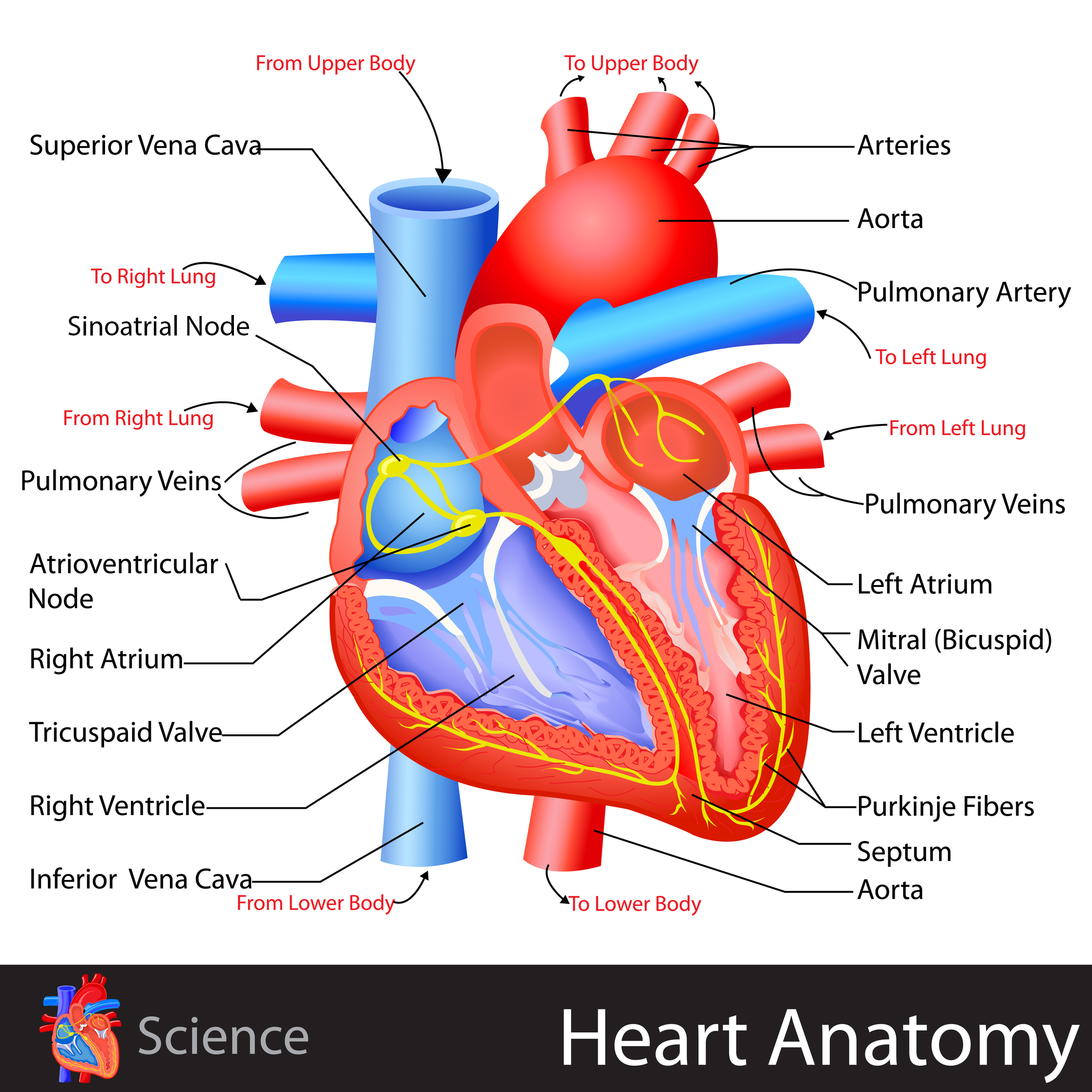
Human Heart Anatomy Diagram Health Images Reference
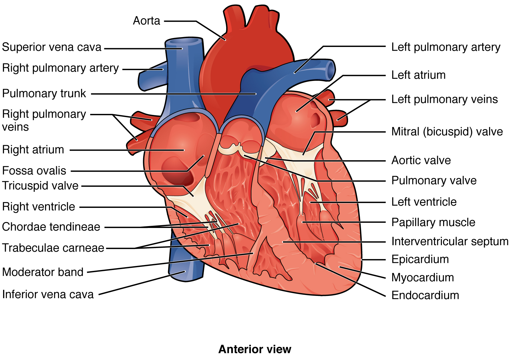
Heart Anatomy · Anatomy and Physiology

Basic Anatomy of the Human Heart Cardiology Associates of Michigan
Heart Anatomy
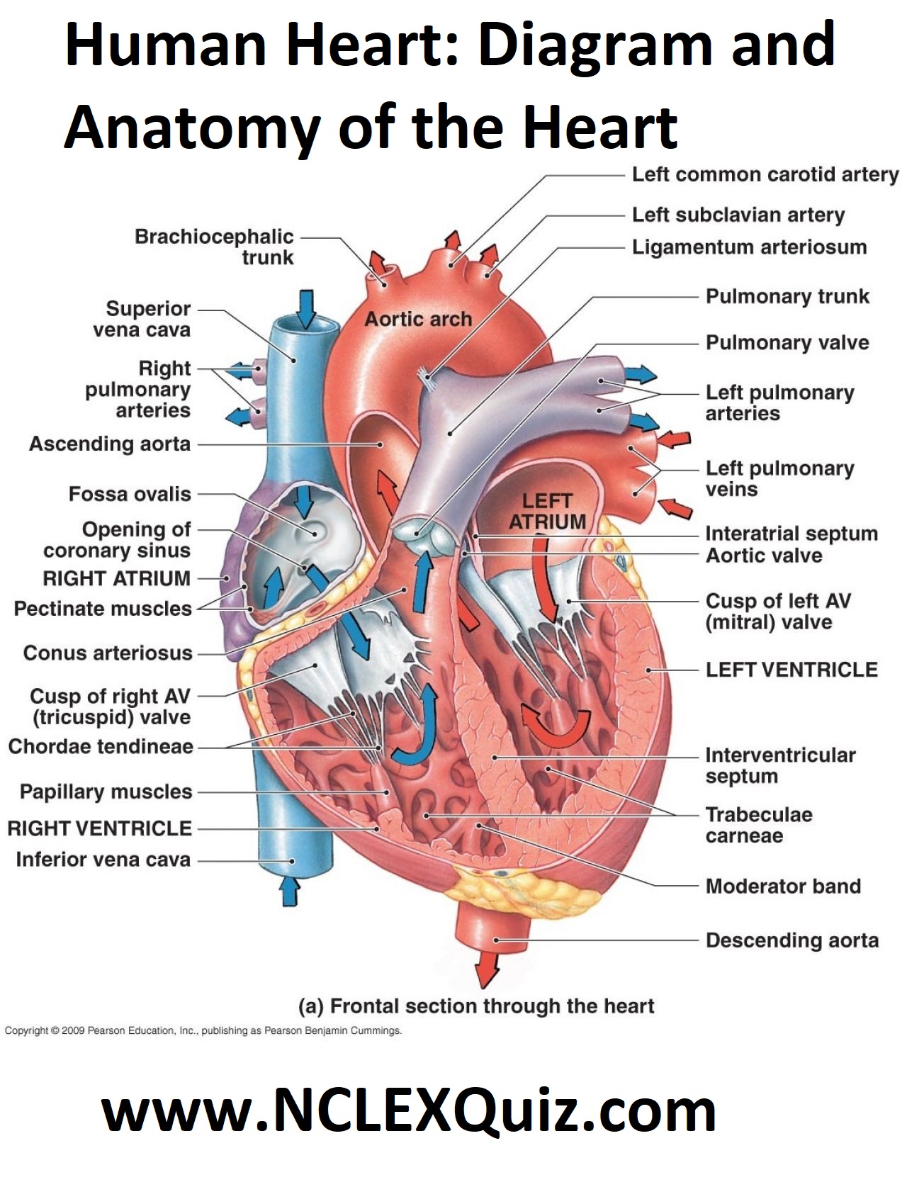
Human Heart Diagram and Anatomy of the Heart StudyPK
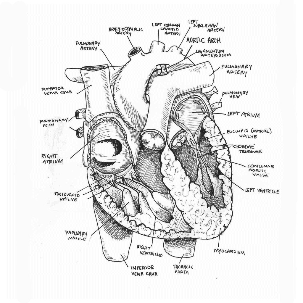
Anatomical Drawing Heart at GetDrawings Free download

External Structure Of Heart Anatomy Diagram

How to Draw the Internal Structure of the Heart (with Pictures)

How to Draw the Internal Structure of the Heart 13 Steps

Heart Anatomy chambers, valves and vessels Anatomy & Physiology
Web Health Library / Body Systems & Organs / Heart Heart Your Heart Is The Main Organ Of Your Cardiovascular System, A Network Of Blood Vessels That Pumps Blood Throughout Your Body.
Cross Sections (Right/Left Ventricle, Right/Left Atrium, Interventricular Septum.
Right, Left, Superior, And Inferior:
The Right Margin Is The Small Section Of The Right Atrium That Extends Between The Superior And Inferior Vena Cava.
Related Post: