Draw And Label Microscope
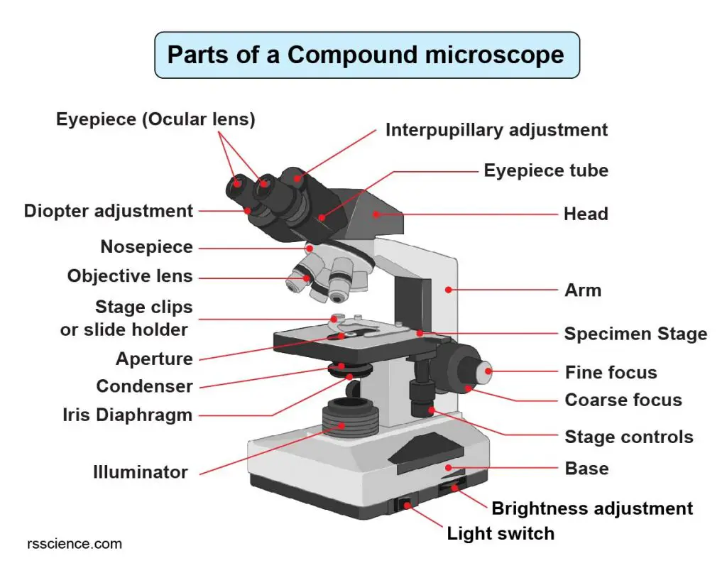
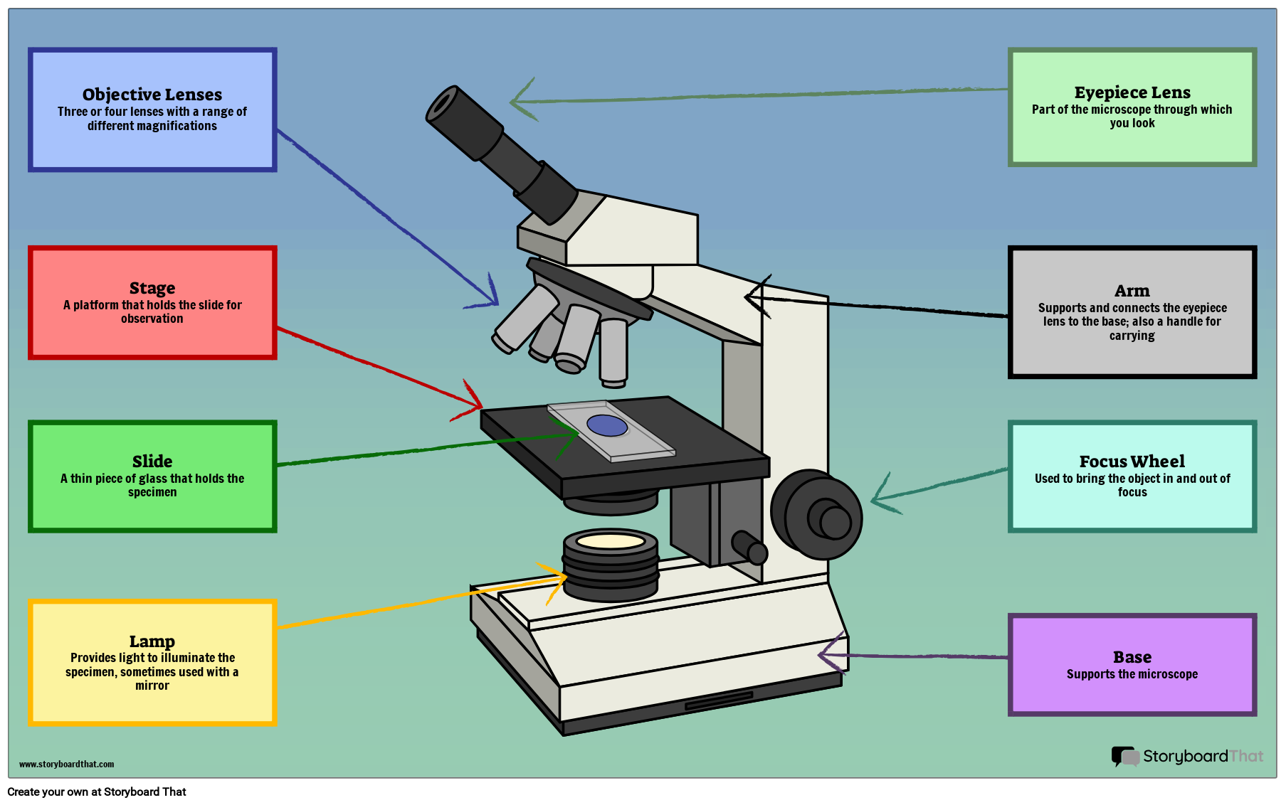
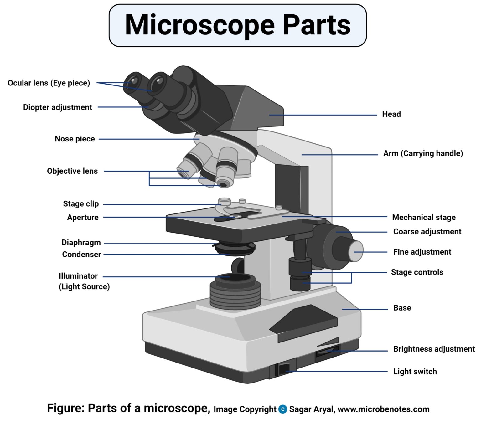
Draw And Label Microscope - In this interactive, you can label the different parts of a microscope. Draw the objective lenses 1.5 step 5: Outline the slide platform 1.6 step 6: Differentiate between a condenser and an abbe condenser. Also indicate the estimated cell size in micrometers under your drawing. Web how to draw a microscope 🔬. Draw the base of the microscope sketch 1.7 step 7: Most photographs of cells are taken using a microscope, and these pictures can also be called micrographs. Today, we're learning how to draw a cool microscope!👩🎨 join our art hub membership! The eyepiece usually contains a 10x or 15x power lens. Provide them with diagrams or actual microscopes, and guide them through the function of each part, such as the lens, eyepiece, and stage. Web a microscope is an instrument that magnifies objects otherwise too small to be seen, producing an image in which the object appears larger. The part that is looked through at the top of the compound microscope.. Web simple microscope is a magnification apparatus that uses a combination of double convex lens to form an enlarged, erect image of a specimen. Web download the label the parts of the microscope pdf printable version here. The part that is looked through at the top of the compound microscope. Label the cell wall, cell membrane, cytoplasm, and chloroplasts in. Web table of contents 1 how to draw a microscope that is hyperrealistic 1.1 step 1: It is categorized into two: Outline the arm frame 1.4 step 4: Web ready to take your drawing skills to the next level? Outline the slide platform 1.6 step 6: Web art for kids hub. Continue follow my channel and like, share,comm. Download the diagrams and practice labeling the different parts of these. Knobs (fine and coarse) 6. The lens the viewer looks through to see the specimen. Review the principles of light microscopy and identify the major parts of the microscope. The eyepiece usually contains a 10x or 15x power lens. And drop the text labels onto the microscope diagram. Web use this interactive to identify and label the main parts of a microscope. Shape the microscope head 1.3 step 3: Label the cell wall, cell membrane, cytoplasm, and chloroplasts in your lab manual. Outline the arm frame 1.4 step 4: Outline the slide platform 1.6 step 6: Knobs (fine and coarse) 6. Describe the functions of each part of the microscope you have drawn above. Useful as a means to change focus on one eyepiece so as to correct for any difference in vision between your two eyes. Compound microscope definitions for labels eyepiece (ocular lens) with or without pointer: We’ll have covered the parts of both simple and compound microscopes and their functions in this article. Web art for kids hub. Review the principles. We’ll have covered the parts of both simple and compound microscopes and their functions in this article. Diagrammatically, identify the various parts of a microscope. Web a labeled diagram of microscope parts furnishes comprehensive information regarding their composition and spatial arrangement within the microscope, enabling researchers to comprehend their function effectively. There are six printables available. Web download the label. There are six printables available. Diagrammatically, identify the various parts of a microscope. We’ll have covered the parts of both simple and compound microscopes and their functions in this article. However, as the saying goes, ‘practice makes perfect’, here is a blank compound microscope diagram and blank electron microscope diagram to label. Outline the slide platform 1.6 step 6: Shape the microscope head 1.3 step 3: Most photographs of cells are taken using a microscope, and these pictures can also be called micrographs. Answers pdf printable version here. And drop the text labels onto the microscope diagram. Outline the slide platform 1.6 step 6: Web pencil drawing paper crayons or colored pencils black marker (optional) draw a microscope printable pdf (see bottom of lesson) the goal is to complete a drawing of a microscope by creating each part one part at a time. Continue follow my channel and like, share,comm. Today, we're learning how to draw a cool microscope!👩🎨 join our art hub membership! Web ready to take your drawing skills to the next level? We’ll have covered the parts of both simple and compound microscopes and their functions in this article. Be sure to indicate the magnification used and specimen name. Differentiate between a condenser and an abbe condenser. Always lift a microscope by holding both the arm and base with two hands. Outline the arm frame 1.4 step 4: Web download the label the parts of the microscope pdf printable version here. Microscope world explains the parts of the microscope, including a printable worksheet for. Knobs (fine and coarse) 6. Web table of contents 1 how to draw a microscope that is hyperrealistic 1.1 step 1: Useful as a means to change focus on one eyepiece so as to correct for any difference in vision between your two eyes. Web a labeled diagram of microscope parts furnishes comprehensive information regarding their composition and spatial arrangement within the microscope, enabling researchers to comprehend their function effectively. Web use this interactive to identify and label the main parts of a microscope.
Compound Microscope Parts Labeled Diagram and their Functions (2023)

36+ Label Each Part Of A Microscope Gif Diagram Printabel

Microscope Drawing And Label at GetDrawings Free download

Compound Light Microscope Drawing at GetDrawings Free download

5 Types of Microscopes with Definitions, Principle, Uses, Labeled Diagrams

Label the Microscope Diagram Download Scientific Diagram

Microscope diagram Tom Butler Science skills, Microscope parts

The Wonders Of Microscopes What You Need To Know Creyentes Diverses News

Microscope Diagram Labeled, Unlabeled and Blank Parts of a Microscope

Parts of a microscope with functions and labeled diagram
Eyepiece (10X) And Objective Lenses (4X, 10X, 40X, 100X).
Web Simple Microscope Is A Magnification Apparatus That Uses A Combination Of Double Convex Lens To Form An Enlarged, Erect Image Of A Specimen.
Learn How To Use The Microscope To View Slides Of Several Different Cell Types, Including The Use Of The Oil Immersion Lens To View Bacterial Cells.
The Part That Is Looked Through At The Top Of The Compound Microscope.
Related Post: