Draw Neuron And Label
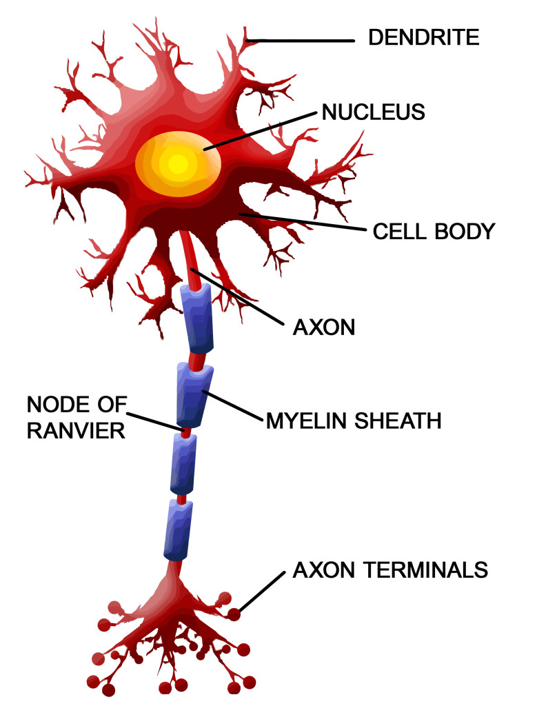
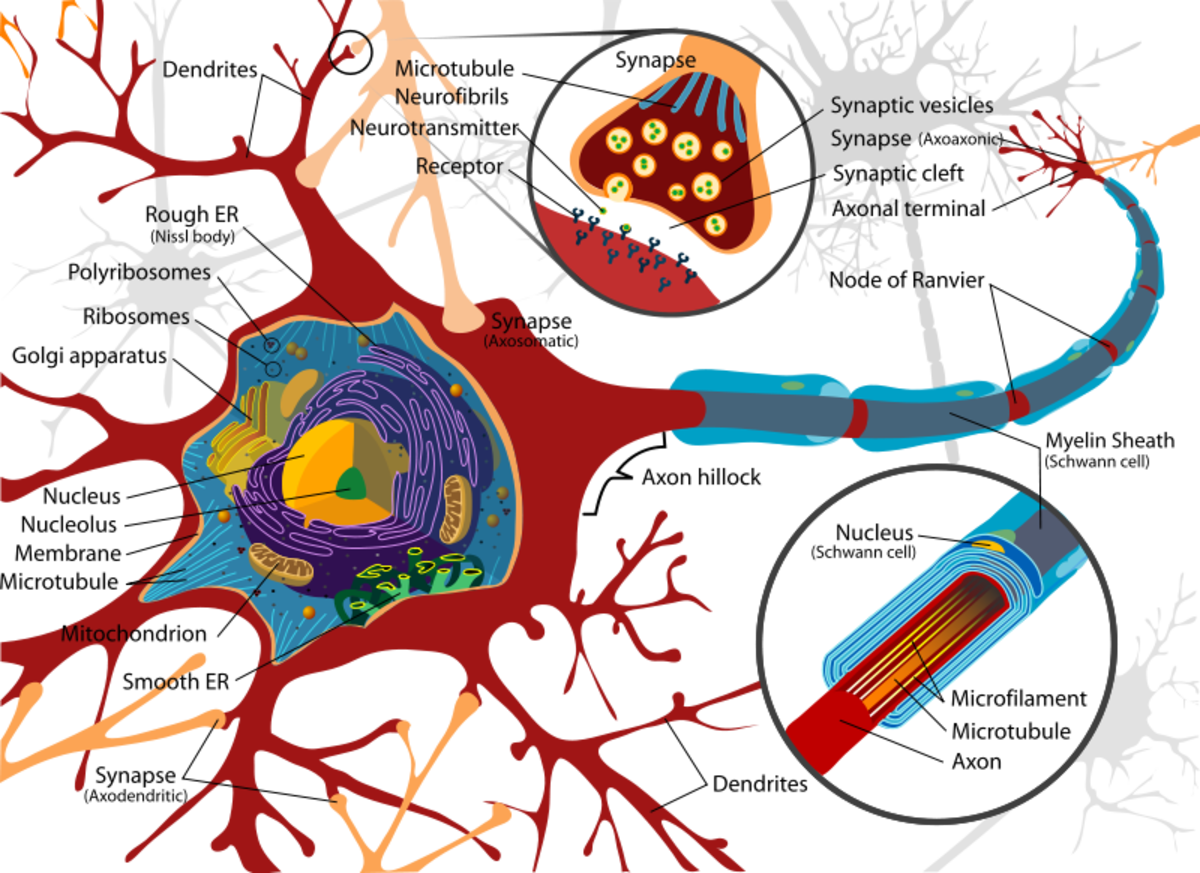
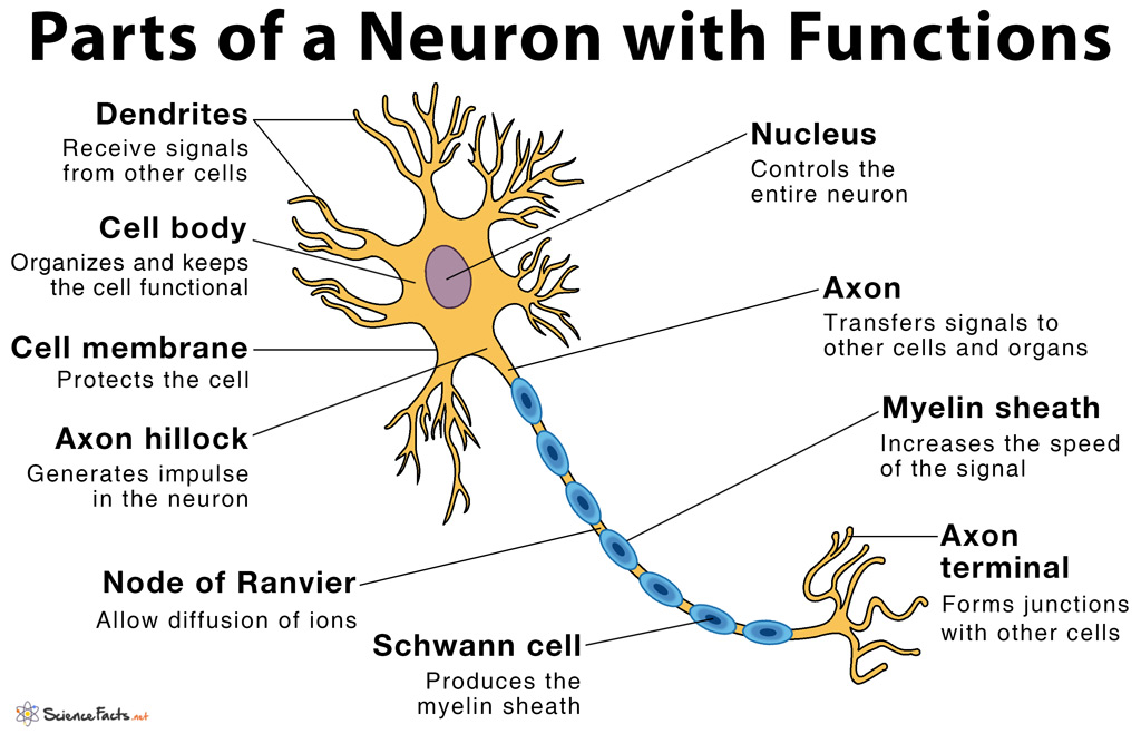
Draw Neuron And Label - Also, the segment of nerve fibre between the two nodes is termed the internode. (10) this neuron part contains instructions for making proteins that. They are the fundamental units of the nervous system and the brain. The region where the myelin sheath is absent is termed the node of ranvier. In these synapses, ions flow directly between cells. What does a neuron look like the main structure of a neuron includes the following parts: What is a membrane potential? A beautiful drawing of a neuron. Web november 9, 2023 reviewed by saul mcleod, phd neurons are the information processing units of the brain responsible for sending, receiving, and transmitting electrochemical signals throughout the body. Watch the video and please be kind enough to thumbs. Web choose the correct names for the parts of the neuron. Signals are received through the dendrites, travel to the cell body, and continue down the axon until they reach the synapse (the communication point between. These carry signals from the central nervous system to other parts of your body such as your muscles and glands. Web draw a neuron,. These synapses communicate using chemical messengers. At the end of the terminal button is a gap known as a synapse. Web choose the correct names for the parts of the neuron. Difference between neurons and neuroglia neuron types there are three different types of neurons: The neuron is a specialized and individual cell, which is also known as the nerve. It is the nutritional center of the neuron where macromolecules are produced. If not, then it’s time to learn! Start studying label parts of a neuron. (8) this neuron part gives messages to muscle tissue. Learn vocabulary, terms, and more with flashcards, games, and other study tools. Neurotransmitters carry signals across the synapse to other neurons. And it will teach you to draw the neuron very easily. Neurons, also known as nerve cells, send and receive signals from your brain. If not, then it’s time to learn! Web neurons are composed of three main parts: What is a membrane potential? A diagram of a neuron also known as the nerve cell is useful as a visual tool to illustrate the various components of the neuron. Web november 9, 2023 reviewed by saul mcleod, phd neurons are the information processing units of the brain responsible for sending, receiving, and transmitting electrochemical signals throughout the body. It. At a synapse, one neuron sends a message to a target neuron—another cell. Signals are received through the dendrites, travel to the cell body, and continue down the axon until they reach the synapse (the communication point between. Difference between neurons and neuroglia neuron types there are three different types of neurons: Label the dendrites, the cell body, and the. Myelin is a thick sheath of lipoprotein that insulates the nerve fibres of the myelinated neurons. At a synapse, one neuron sends a message to a target neuron—another cell. They are responsible for the electrical signals that communicate information about sensations, and that produce movements in response to those stimuli, along with inducing thought processes within the brain. What is. Is membrane potential always the same along the entire neuron? A neuron is a cell in the brain that processes information. These carry signals from the central nervous system to other parts of your body such as your muscles and glands. Web multipolar neurons have one axon and many dendritic branches. What does a neuron look like the main structure. Neurons, also known as nerve cells, are essentially the cells that make up the brain and the nervous system. (10) this neuron part contains instructions for making proteins that. A group of neurons forms a nerve. It is not a continuous sheath and is absent at regular intervals. (8) this neuron part gives messages to muscle tissue. Spreads the cell's impulse out to reach other neurons. Label the dendrites, the cell body, and the axon. These synapses communicate using chemical messengers. Neurons, also known as nerve cells, send and receive signals from your brain. Web neurons communicate with one another at junctions called synapses. (6) this neuron part receives messages from other neurons. Spreads the cell's impulse out to reach other neurons. Label the dendrites, the cell body, and the axon. Web carries the cell's impulse to the terminal. The neuron is a specialized and individual cell, which is also known as the nerve cell. Web terminal buttons are found at the end of the axon, below the myelin sheath, and are responsible for sending the signal on to other neurons. Start studying label parts of a neuron. Web the neuron is the one that transfers the message (nerve impulse) to the brain. A beautiful drawing of a neuron. In these synapses, ions flow directly between cells. Indicate the direction in which signals typically travel along the neuron. Why do you need to know this? Neurons are the cells considered to be the basis of nervous tissue. So about taste sensation, the stimuli is actually the chemicals in our mouth. Web neurons communicate with one another at junctions called synapses. What is a membrane potential?
The Neuron External Structure and Classification Interactive

draw a typical multipolar neuron in the space below

A simple guide to neuron diagrams and types (2023)

Neuron Diagram Straight from a Scientist

FileNeuron1.jpg Wikimedia Commons

Structure of a Neuron Owlcation

Draw A Labelled Diagram Showing The Structure Of A Neuron Biology

How TO Draw neuron step by step easy/diagram of neuron/neuron drawing

Anatomy Of A Neuron ANATOMY

Neuron Study Guide Inspirit Learning Inc
Web Parts Of A Neuron And Their Functions With Labelled Diagram Parts Of A Neuron And Their Function Neurons Or Nerve Cells Are The Basic Building Blocks Or Units Of The Nervous System.
Web How To Draw A Neuron Step By Step For Beginners.
Cell Body, Dendrites, Axon, Synaptic Terminals, Axon Hillock, Myelin Sheath (Aka Myelinated Internode) Nodes Of Ranvier See Above Picture.
They’re Located All Over The Place But Are Mostly Found In The Cortex And Cerebellum.
Related Post: