Drawing Of Esophagus
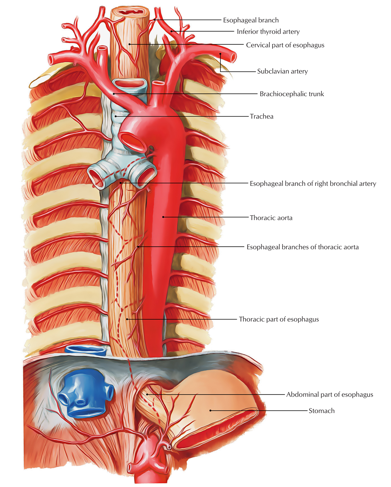
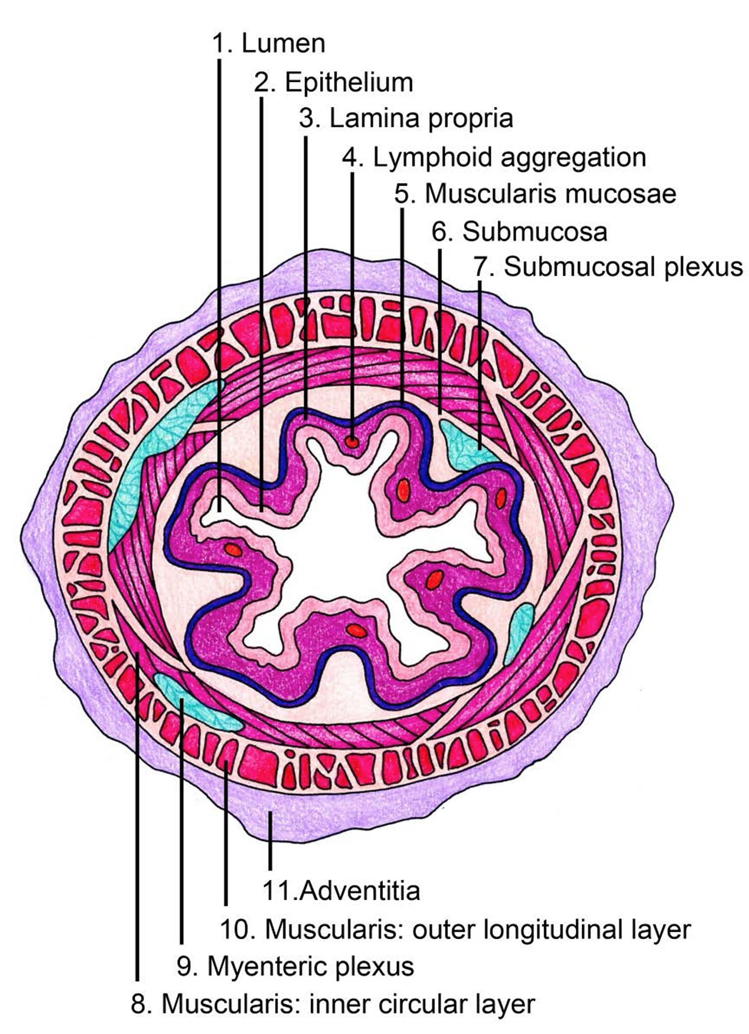
Drawing Of Esophagus - Lamina propria of mucosa layers of esophagus #3. Web choose from drawing of the esophagus stock illustrations from istock. There are three layers of the mucosa: The esophagus is a hollow muscular tube that transports saliva, liquids, and foods from the mouth to the stomach. It forms an important piece of the gastrointestinal tract and functions as the conduit for food and liquids that have been swallowed into. The order of these layers from the inside out are: Drawing of the gi tract, with the esophagus, stomach, small intestine, duodenum, jejunum, ileum, large intestine, cecum, colon, rectum, and anus labeled. Voice box, also known as the larynx; When the patient is upright, the esophagus is usually between 25 to 30. Web medical illustration engraving from 1872 featuring the human digestive tract healthy throat linear icon. Upper section of alimentary canal. One of the most common symptoms of esophagus problems is heartburn, a burning sensation in the middle of your chest. In its abdominal course, it is covered with the peritoneum of the greater sac anteriorly and on its left side, and it is covered with. The order of these layers from the inside out are:. Lymhatic nodules (not in all amimals) oin lamina propria layer #4. Let us learn how to draw esophagus for step by step. Web the esophagus is a muscular channel that carries food from the pharynx to the stomach. The esophagus lies posterior to the trachea and the heart and passes through the mediastinum and the hiatus, an opening in the. It forms an important piece of the gastrointestinal tract and functions as the conduit for food and liquids that have been swallowed into. When food enters the mouth, it mixes with saliva. Different layers of the esophagus. Oral cavity, pharynx, esophagus in good health. One of the most common symptoms of esophagus problems is heartburn, a burning sensation in the. Stratified squamous epithelium on mucosa (keratinized or nonkeratinized; Web the esophagus is a muscular tube about ten inches (25 cm.) long, extending from the hypopharynx to the stomach. Web the esophagus is a long, thin, and muscular tube that connects the pharynx (throat) to the stomach. These layers are similar all throughout the whole digestive tract. The esophagus is a. Problems with the esophagus include acid reflux and gerd. Web the esophagus is a muscular tube about ten inches (25 cm.) long, extending from the hypopharynx to the stomach. Oral cavity, pharynx, esophagus in good health. The throat includes the esophagus; In its abdominal course, it is covered with the peritoneum of the greater sac anteriorly and on its left. Web your esophagus is a hollow, muscular tube that carries food and liquid from your throat to your stomach. The throat includes the esophagus; Editable stroke healthy throat linear icon. Web the esophagus is a muscular tube about ten inches (25 cm.) long, extending from the hypopharynx to the stomach. These layers are similar all throughout the whole digestive tract. Web the esophagus is a long, thin, and muscular tube that connects the pharynx (throat) to the stomach. Oral cavity, pharynx, esophagus in good health. Voice box, also known as the larynx; In its abdominal course, it is covered with the peritoneum of the greater sac anteriorly and on its left side, and it is covered with. These layers are. Web subscribe now : Web anatomic drawings of the digestive system esophagus liver (right lobe) intrahepatic bile duct common bile duct gallbladder duodenum hepatic flexure ascending colon ileum ileocecal valve cecum appendix liver esophageal sphincter liver (left lobe) stomach greater curvature pancreas splenic flexure jejunum descending colon. Muscles in your esophagus propel food down to your stomach. Web your esophagus. Lamina propria of mucosa layers of esophagus #3. Web your esophagus is a hollow, muscular tube that carries food and liquid from your throat to your stomach. Web the esophagus is a muscular tube about ten inches (25 cm.) long, extending from the hypopharynx to the stomach. Different layers of the esophagus. Mayo clinic does not endorse companies or products. Web the esophagus is a muscular channel that carries food from the pharynx to the stomach. These layers are similar all throughout the whole digestive tract. It forms an important piece of the gastrointestinal tract and functions as the conduit for food and liquids that have been swallowed into. Web the esophagus is a long, thin, and muscular tube that. Web the abdominal part of the esophagus. Mayo clinic does not endorse companies or products. The order of these layers from the inside out are: Windpipe, also known as the trachea; It starts with the upper esophageal sphincter, formed in part by the cricopharyngeus muscle, and ends with the lower esophageal sphincter, surrounded by the crural diaphragm. Web subscribe now : When the patient is upright, the esophagus is usually between 25 to 30. The esophagus passes through the right crus of the diaphragm. Drawing of the gi tract, with the esophagus, stomach, small intestine, duodenum, jejunum, ileum, large intestine, cecum, colon, rectum, and anus labeled. Web the esophagus is made up of four layers of tissue. The throat includes the esophagus; Web choose from drawing of the esophagus stock illustrations from istock. Web your esophagus is a hollow, muscular tube that carries food and liquid from your throat to your stomach. The esophagus lies posterior to the trachea and the heart and passes through the mediastinum and the hiatus, an opening in the diaphragm, in its descent from the thoracic to the abdominal cavity. Web simple drawing of anterior view of the arch of the aorta and the many branches of arteries which arise from the thoracic aorta to provide arterial blood supply to the trachea and esophagus. Esophagus drawing stock photos are available in a variety of sizes and formats to fit your needs.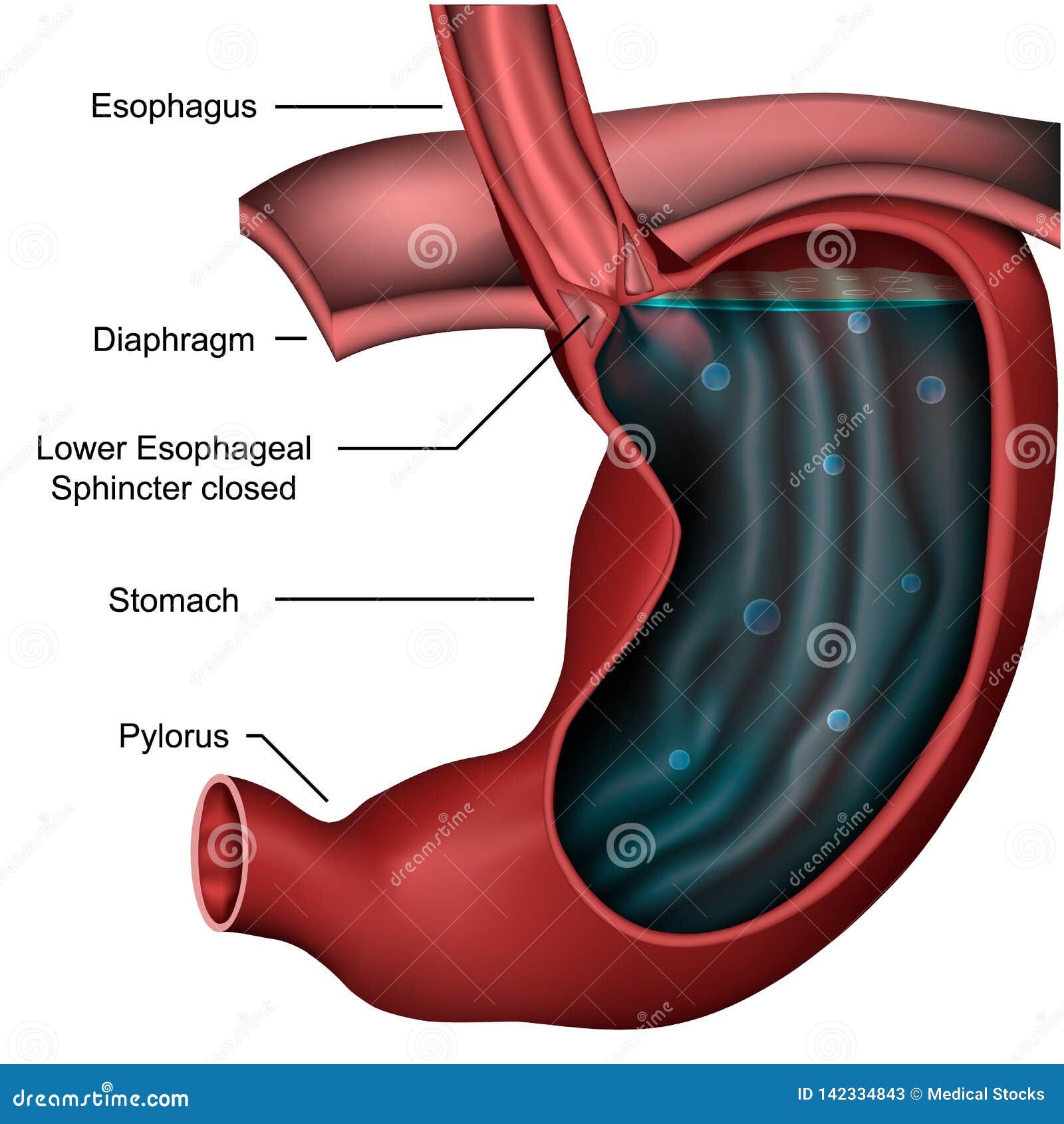
Esophageal Sphincter Anatomy 3d Medical Illustration on White

Anatomy Of The Esophagus

Esophagus / Endoscopy — High Plains Surgical Associates

Esophagus stock illustration. Illustration of throat 40419397
Esophagus Earth's Lab

E.3. Esophagus
![]()
Esophagus Anatomy, sphincters, arteries, veins, nerves Kenhub
The Human Esophagus Functions and Anatomy and Problems

Esophagus Function In The Digestive System
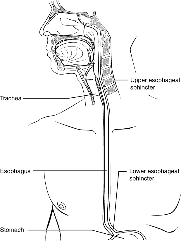
The Mouth, Pharynx, and Esophagus · Anatomy and Physiology
Lamina Propria Of Mucosa Layers Of Esophagus #3.
Web Browse 1,989 Esophagus Anatomy Photos And Images Available, Or Start A New Search To Explore More Photos And Images.
Connective Tissue Papillae Of Esophagus #5.
Web The Esophagus (Oesophagus) Is A 25 Cm Long Fibromuscular Tube Extending From The Pharynx (C6 Level) To The Stomach (T11 Level).
Related Post: