Drawing Of Heart And Lungs
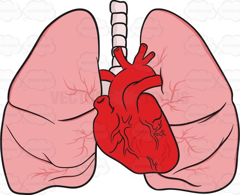
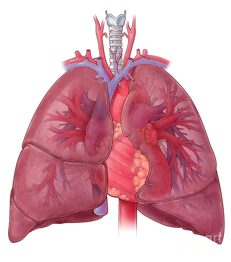
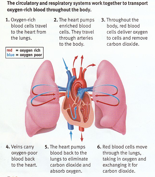
Drawing Of Heart And Lungs - The aia rendering of a chest ct scan reconstructed in 3d shows the lung and heart beyond the skin. It projects upwards, above the level of the 1st rib and into the floor of the neck. Web anatomy of the interior of the heart. Passage of blood through the left atrium, bicuspid valve, left ventricle, aorta , tissues of the body, and back to the right atrium constitutes the systemic circulation. Most popular organs line icons. Web the lungs are roughly cone shaped, with an apex, base, three surfaces and three borders. Web 3d ct of lungs and heart. It transports deoxygenated blood to the lungs to absorb oxygen and release carbon dioxide. Web the heart lung diagram is a visual representation of the relationship between the heart and lungs. Brain, kidney, heart, liver, stomach. The bottom tip of the heart, known as its apex, is turned to the left, so that about 2/3 of the heart is located on the body’s left side with the other 1/3 on right. Heart and lung diagram this image of the heart and lungs is taken from a middle school health textbook. Save time with a video! This. Most popular organs line icons. Heart lungs brain human heart lungs brain heart lungs heart lungs vector heart lungs not blood heart lungs and chest sort by: Passage of blood through the left atrium, bicuspid valve, left ventricle, aorta , tissues of the body, and back to the right atrium constitutes the systemic circulation. Web browse 1,000+ heart and lungs. Web the lungs are roughly cone shaped, with an apex, base, three surfaces and three borders. Within the mediastinum, the heart is separated from the other mediastinal structures by a tough membrane known as the pericardium, or pericardial sac. Web 3d ct of lungs and heart. It projects upwards, above the level of the 1st rib and into the floor. Brain, kidney, heart, liver, stomach. This image shows the four chambers of the heart and the direction that blood flows through the heart. Every breath you take pulls oxygen into your lungs to supply. The beautiful color provides a quiet and restful ambiance as the heart continues its quiet rhythms among the mostly silent and life sustaining movement of air. The heart is responsible for pumping blood throughout the body, while the lungs are responsible for oxygenating that blood. Most popular organs line icons. On the other hand, if your lungs drawing is too large, it could be a sign of overinflation, which could also be a sign of a respiratory condition. Heart lungs brain human heart lungs brain heart. Web the right atrium and ventricle receive deoxygenated blood from systemic veins and pump it to the lungs, while the left atrium and ventricle receive oxygenated blood from the lungs and pump it to the systemic vessels which distribute it throughout the body. On the other hand, if your lungs drawing is too large, it could be a sign of. Web the human heart is located within the thoracic cavity, medially between the lungs in the space known as the mediastinum. Brain, kidney, heart, liver, stomach. Most popular organs line icons. The full page where it appears can be seen below. Web the right atrium and ventricle receive deoxygenated blood from systemic veins and pump it to the lungs, while. Web anatomy of the interior of the heart. How to draw lungs | human organs drawing | easy step by step drawing for kids subscribe for more videos: To zoom, making this image accessible this image can be made accessible with an image description or by using a tactile graphic. Web the human heart is located within the thoracic cavity,. It projects upwards, above the level of the 1st rib and into the floor of the neck. Heart lungs brain human heart lungs brain heart lungs heart lungs vector heart lungs not blood heart lungs and chest sort by: Structures and blood flow [cardiology made easy] anatomy of the human heart made easy using labeled diagrams of the main cardiac. On the other hand, if your lungs drawing is too large, it could be a sign of overinflation, which could also be a sign of a respiratory condition. Figure 19.2 shows the position of the heart within the thoracic cavity. The heart is responsible for pumping blood throughout the body, while the lungs are responsible for oxygenating that blood. Within. Web the heart is a muscular pumping organ located medial to the lungs along the body’s midline in the thoracic region. Every breath you take pulls oxygen into your lungs to supply. Web browse 1,000+ heart and lungs drawings stock photos and images available, or start a new search to explore more stock photos and images. The beautiful color provides a quiet and restful ambiance as the heart continues its quiet rhythms among the mostly silent and life sustaining movement of air in the airways. To zoom, making this image accessible this image can be made accessible with an image description or by using a tactile graphic. Within the mediastinum, the heart is separated from the other mediastinal structures by a tough membrane known as the pericardium, or pericardial sac. Brain, kidney, heart, liver, stomach. This image shows the four chambers of the heart and the direction that blood flows through the heart. Heart and lung diagram this image of the heart and lungs is taken from a middle school health textbook. Passage of blood through the left atrium, bicuspid valve, left ventricle, aorta , tissues of the body, and back to the right atrium constitutes the systemic circulation. It transports deoxygenated blood to the lungs to absorb oxygen and release carbon dioxide. It projects upwards, above the level of the 1st rib and into the floor of the neck. The heart is responsible for pumping blood throughout the body, while the lungs are responsible for oxygenating that blood. Web the lungs are roughly cone shaped, with an apex, base, three surfaces and three borders. Heart lungs brain human heart lungs brain heart lungs heart lungs vector heart lungs not blood heart lungs and chest sort by: Figure 19.2 shows the position of the heart within the thoracic cavity.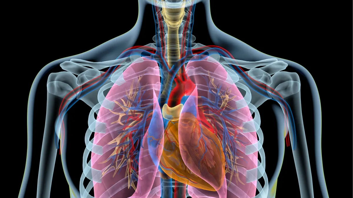
The heart & the lungs What's the connection? Blog ndd Medical

Lungs and human heart illustration Stock vector Colourbox
Heart And Lungs Drawing at GetDrawings Free download

Anatomy of the Heart and Lungs Diagnosis 101
Heart And Lung Anatomy, Illustration Photograph by Evan Oto Pixels

Anatomy of the Heart and Lungs with Pulmonary Artery Circulation

Diagram showing heart and lungs blood flow illustration Stock Vector
Sample 1 Heart and Lung Diagram DIAGRAM Center
19th Century Medical Engraving Of Human Lungs And Heart Stock Vector

Heart And Lung Anatomy, Illustration by Evan Oto in 2021 Lung anatomy
Web The Heart Lung Diagram Is A Visual Representation Of The Relationship Between The Heart And Lungs.
The Oxygenated Blood Then Flows Back To The Heart.
Web Anatomy Cardiology Feb 24 Anatomy Of The Heart:
Most Popular Human Organs Hand Drawn Line Icon Set Set Of Isolated Internal Human Organ Sketch Set Of Isolated Internal Human Organ Sketch.
Related Post:
