Golgi Complex Drawing
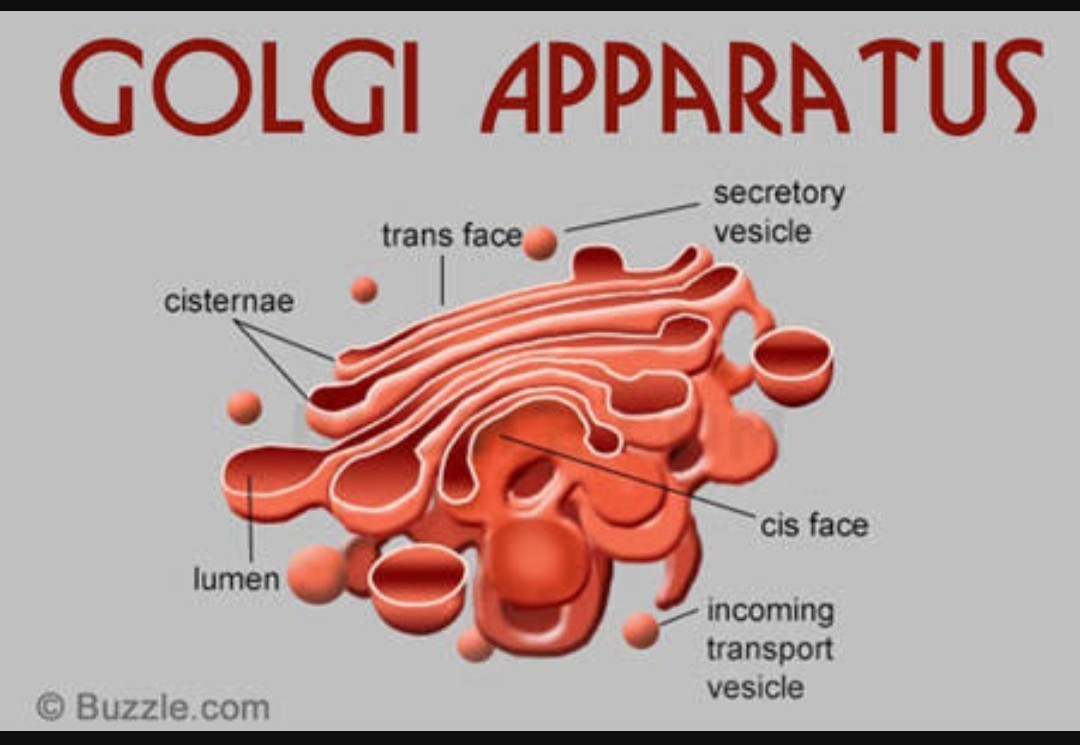
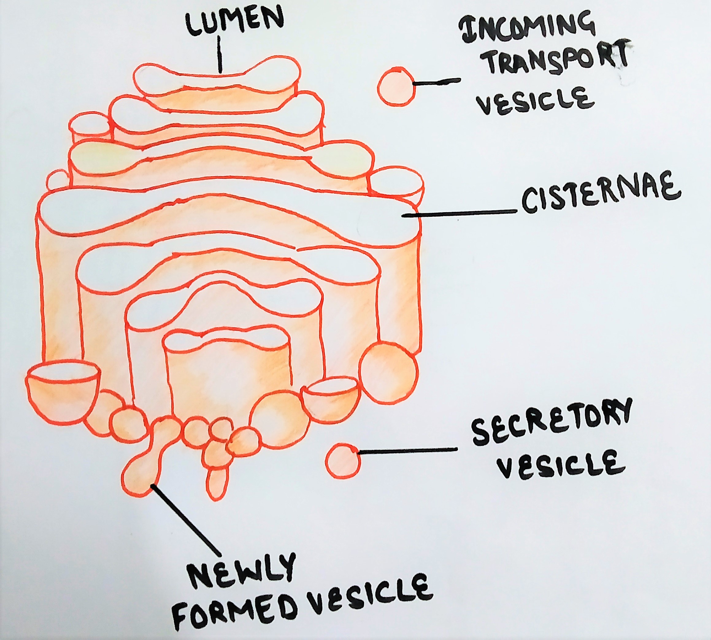
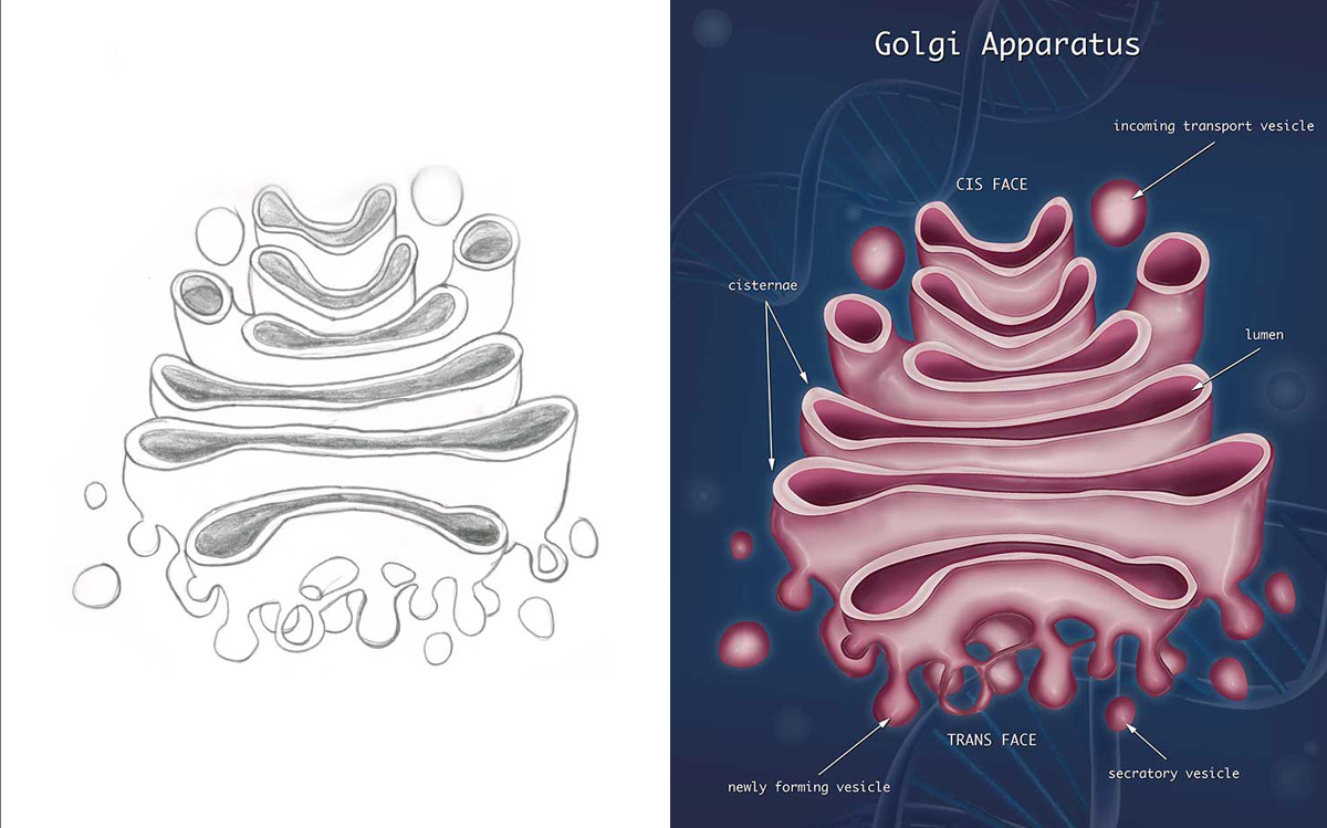
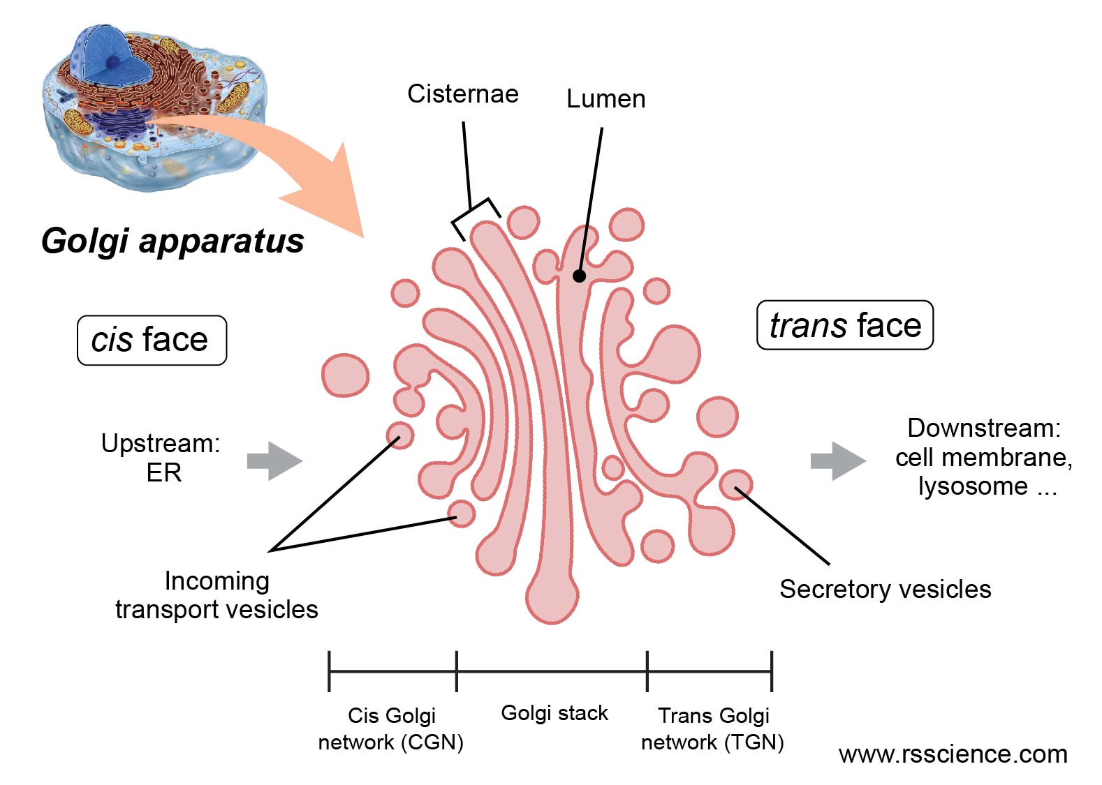
Golgi Complex Drawing - Golgi's method is a silver staining technique that is used to visualize nervous tissue under light microscopy. This intracellular structure is universally known nowadays as “golgi apparatus”. In fact, even though the golgi was first seen in 1897, scientists are still working on a model that. | structure of golgi complex | easy step by step diagram | biology bise sciences 2.2k subscribers subscribe 39 2.7k views 3 years ago uploaded videos it's an. In addition, as noted earlier, glycolipids and sphingomyelin are synthesized within the golgi. This cytoplasmic organelle is named after its discoverer golgi. Lysosomes, the plasma membrane, or secretion. The number of ‘golgi apparatus’ within a cell is variable. The golgi body is no doubt a complex and a ripe area for ongoing research. Web so, the golgi apparatus, number one, modifies proteins that are made in the rough endoplasmic reticulum. The structure was discovered in 1898. The name “golgi complex” was given after its discoverer, camilo golgi (1898), an italian anatomist. The number of ‘golgi apparatus’ within a cell is variable. Web the golgi apparatus, sometimes called the golgi complex or golgi body, is responsible for manufacturing, warehousing, and shipping certain cellular products, particularly those from the endoplasmic reticulum (er).. Web this drawing shows how the rough endoplasmic reticulum forms vesicles (without ribosomes attached) that carry the newly. Structure of golgi complex 3. A complex network of tubules and vesicles is located at the edges of the cisternae. Cisternae, vesicles and vacuoles are the three major darts of golgi complex (fig. Web so, the golgi apparatus, number one, modifies proteins. Web the golgi apparatus is comprised of a series of flattened sacs that extend from the endoplasmic reticulum. Animal cells tend to have fewer and larger golgi apparatus. Modify certain proteins and glycoproteins; Web the golgi apparatus, also known as the golgi complex, golgi body, or simply the golgi, is an organelle found in most eukaryotic cells. Web drawing of. Italian histologist camillo golgi (1898) first discovered golgi apparatus in the nerve cells of. The golgi complex functions to: Sort proteins and lipids received from the er; The cisternae contain enzymes that modify proteins passing through them. The name “golgi complex” was given after its discoverer, camilo golgi (1898), an italian anatomist. | structure of golgi complex | easy step by step diagram | biology bise sciences 2.2k subscribers subscribe 39 2.7k views 3 years ago uploaded videos it's an. Web the golgi apparatus, or golgi complex, functions as a factory in which proteins received from the er are further processed and sorted for transport to their eventual destinations: Structure of golgi. Web the golgi apparatus, sometimes called the golgi complex or golgi body, is responsible for manufacturing, warehousing, and shipping certain cellular products, particularly those from the endoplasmic reticulum (er). The golgi complex functions to: It is also called golgi complex, golgi bodies, golgi some, golgi membrane, dictyosomes, lipochondria, dalton complex, baker’s bodies, carbohydrate factory, ‘traffic police’ of cell etc. And. Italian histologist camillo golgi (1898) first discovered golgi apparatus in the nerve cells of. This cytoplasmic organelle is named after its discoverer golgi. Web this drawing shows how the rough endoplasmic reticulum forms vesicles (without ribosomes attached) that carry the newly. The number of ‘golgi apparatus’ within a cell is variable. The golgi body is no doubt a complex and. Sort proteins and lipids received from the er; Depending on the type of cell, there can be just a few complexes or there can be hundreds. This cytoplasmic organelle is named after its discoverer golgi. Web [in this figure] a 3d drawing of the golgi apparatus. Animal cells tend to have fewer and larger golgi apparatus. Web the golgi apparatus, sometimes called the golgi complex or golgi body, is responsible for manufacturing, warehousing, and shipping certain cellular products, particularly those from the endoplasmic reticulum (er). This cytoplasmic organelle is named after its discoverer golgi. Web so, the golgi apparatus, number one, modifies proteins that are made in the rough endoplasmic reticulum. And number three, the golgi. Modify certain proteins and glycoproteins; | structure of golgi complex | easy step by step diagram | biology bise sciences 2.2k subscribers subscribe 39 2.7k views 3 years ago uploaded videos it's an. Web the golgi apparatus, or golgi complex, functions as a factory in which proteins received from the er are further processed and sorted for transport to their. Web 42k views 3 years ago #drawing. Web the golgi apparatus, or golgi complex, functions as a factory in which proteins received from the er are further processed and sorted for transport to their eventual destinations: Golgi's method is a silver staining technique that is used to visualize nervous tissue under light microscopy. Web golgi’s drawings of the “internal reticular apparatus” that he observed in spinal ganglia (the different drawings illustrate the variety of features golgi observed with his metal impregnation, from opera omnia). Web the golgi’s function is an ongoing mystery. In fact, even though the golgi was first seen in 1897, scientists are still working on a model that. Web the golgi apparatus is comprised of a series of flattened sacs that extend from the endoplasmic reticulum. It is also called golgi complex, golgi bodies, golgi some, golgi membrane, dictyosomes, lipochondria, dalton complex, baker’s bodies, carbohydrate factory, ‘traffic police’ of cell etc. Web the golgi apparatus, also known as the golgi complex, golgi body, or simply the golgi, is an organelle found in most eukaryotic cells. Web so, the golgi apparatus, number one, modifies proteins that are made in the rough endoplasmic reticulum. How to draw golgi apparatus/golgi body structure of function, how to draw golgi body easy/diagram of golgi body/golgi body drawing, how to draw golgi apparatus. Depending on the type of cell, there can be just a few complexes or there can be hundreds. Golgi bodies are found in almost all the animal and plant cells. Structure of golgi complex 3. This cytoplasmic organelle is named after its discoverer golgi. Modify certain proteins and glycoproteins;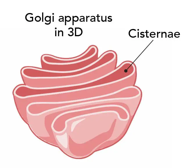
Golgi Apparatus Function the Post Office inside the Cells Rs' Science

Structure and function of the golgi apparatus

Golgi apparatus detailed illustration Illustration, Vector images, Vector
Golgi Apparatus Drawing at Explore collection of Golgi Apparatus Drawing
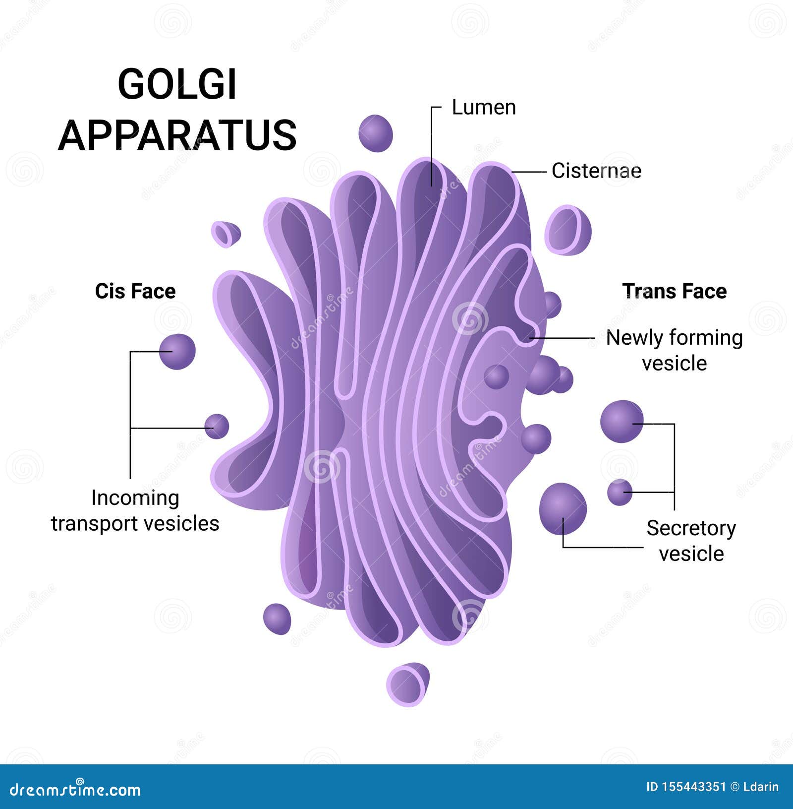
Illustration of the Golgi Apparatus Structure. Vector Infographics Stock Vector Illustration

Golgi apparatus physiology
Golgi complex and its role in protein secretion The Virtual Notebook
Golgi Apparatus Sketch at Explore collection of Golgi Apparatus Sketch
Golgi Apparatus Function the Post Office inside the Cells Rs' Science

how to draw diagram of golgi body easily, YouTube
Web This Drawing Shows How The Rough Endoplasmic Reticulum Forms Vesicles (Without Ribosomes Attached) That Carry The Newly.
Web [In This Figure] A 3D Drawing Of The Golgi Apparatus.
Web In This Video, We Delve Into The Inner Workings Of A Vital Cellular Organelle, The Golgi Apparatus, Also Know As Golgi Complex And Golgi Body.
In Addition, As Noted Earlier, Glycolipids And Sphingomyelin Are Synthesized Within The Golgi.
Related Post: