Knee Joint Drawing
Knee Joint Drawing - Web arthrocentesis of the knee is the process of puncturing the knee (tibiofemoral and patellofemoral) joint with a needle to withdraw synovial fluid. Rheumatology and orthopedics traumatology medicine banners. To understand the function and structure of the knee joint, a knee anatomy can be helpful. We need to identify four bony bits to draw the knee well. How much fluid is typically drained from the knee Four quadriceps muscles are present in front of the knee which help in straightening the leg from the knee. Web the muscles that affect the knee’s movement run along the thigh and calf. (see also evaluation of the patient with joint symptoms and evaluation of the knee.) indications for knee arthrocentesis diagnosis of the cause of a synovial effusion (eg, infection Web in this video we'll show you how to draw a knee. Anatomy where is the knee joint located? Anatomy sketch current as of: To understand the function and structure of the knee joint, a knee anatomy can be helpful. It is a surgical procedure that involves draining the knee joint, also known as arthrocentesis. They are attached to the femur (thighbone), tibia (shinbone), and fibula (calf bone) by fibrous tissues called ligaments. Its complex structure and constant utilization. Knee anatomy involves more than just muscles and bones. Four quadriceps muscles are present in front of the knee which help in straightening the leg from the knee. Web find a location find a physician knee overview as one of the largest and most advanced joints in the human body, the knee is critical to mobility. The tibiofemoral joint and. The tibiofemoral joint is an articulation between the tibia and the femur, while the patellofemoral joint is an articulation between the patella. Web ⚫ muscles quadriceps and hamstring muscles are the major ones which are associated with the knee joint. Web there are four main movements that the knee joint permits: The knee is the largest joint, and its primary. Web find a location find a physician knee overview as one of the largest and most advanced joints in the human body, the knee is critical to mobility. Web in this video we'll show you how to draw a knee. Web anatomy of the knee. The knee is the meeting point of the femur (thigh bone) in the upper leg. To understand the function and structure of the knee joint, a knee anatomy can be helpful. They are attached to the femur (thighbone), tibia (shinbone), and fibula (calf bone) by fibrous tissues called ligaments. Side and front view of knee bones, hand drawn femur, patella, tibia and fibula, tibial plateau and lateral condyle. Pause as you need to and practice. Four quadriceps muscles are present in front of the knee which help in straightening the leg from the knee. Web anatomy explorer adductor tubercle anterior cruciate ligament articular cartilage (femur) articular cartilage (tibia) femur fibula infrapatellar fat pad interosseous membrane of the leg joint capsule of knee lateral collateral (fibular collateral) ligament lateral condyle of femur lateral condyle of tibia. They are attached to the femur (thighbone), tibia (shinbone), and fibula (calf bone) by fibrous tissues called ligaments. Web find a location find a physician knee overview as one of the largest and most advanced joints in the human body, the knee is critical to mobility. Web how do i use knee drainage? Web ⚫ muscles quadriceps and hamstring muscles. Web function what does the knee joint do? Knee joint isolated vector illustration, flat design. Find out how the joint fits together in our knee anatomy diagram and what goes wrong. (see also evaluation of the patient with joint symptoms and evaluation of the knee.) indications for knee arthrocentesis diagnosis of the cause of a synovial effusion (eg, infection Web. Web there are four main movements that the knee joint permits: The femur, tibia and patella. Web anatomy explorer adductor tubercle anterior cruciate ligament articular cartilage (femur) articular cartilage (tibia) femur fibula infrapatellar fat pad interosseous membrane of the leg joint capsule of knee lateral collateral (fibular collateral) ligament lateral condyle of femur lateral condyle of tibia lateral epicondyle of. Knee joint icon gray illustration on white. The femur, tibia and patella. The knee is the meeting point of the femur (thigh bone) in the upper leg and. Find out how the joint fits together in our knee anatomy diagram and what goes wrong. To understand the function and structure of the knee joint, a knee anatomy can be helpful. Web there are four main movements that the knee joint permits: Web anatomy of human knee vector sketch of leg bones and joint, medicine design. How much fluid is typically drained from the knee Ligaments, tendons, and cartilage work together to connect the thigh bone, shin bone, and knee cap and allow the leg to bend back and forth like a hinge. Anatomy where is the knee joint located? The (1) femur, extending from the hip to the knee, the (2) tibia extending from the knee to the foot, the (3) fibula following along the tibia on the outer side of the leg, and the (4) patella or the kneecap protecting the knee joint. Knee joint isolated vector illustration, flat design. The knee is the largest joint, and its primary function is helping in mobility. It is a complex hinge joint composed of two articulations; The hamstring muscles are present at the back of the knee and help in bending. Its complex structure and constant utilization causes it to be particularly vulnerable to damage. It is a surgical procedure that involves draining the knee joint, also known as arthrocentesis. The femur, tibia and patella. The tibiofemoral joint is an articulation between the tibia and the femur, while the patellofemoral joint is an articulation between the patella. Web the knee joint is a synovial joint that connects three bones; Web how do i use knee drainage?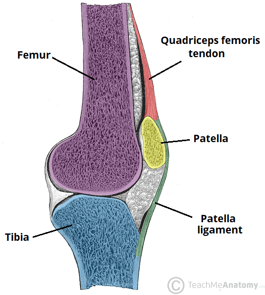
The Knee Joint Articulations Movements Injuries TeachMeAnatomy
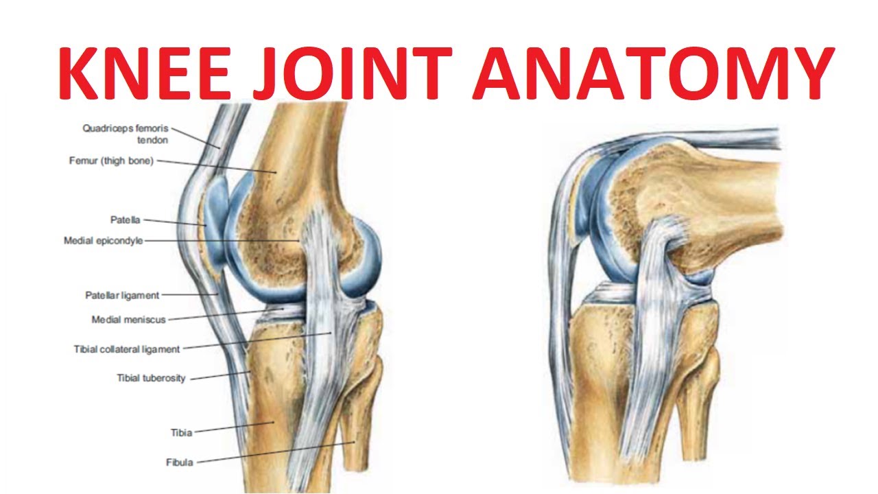
Knee Joint Anatomy YouTube
Knee Joint Showing Interior Ligaments ClipArt ETC
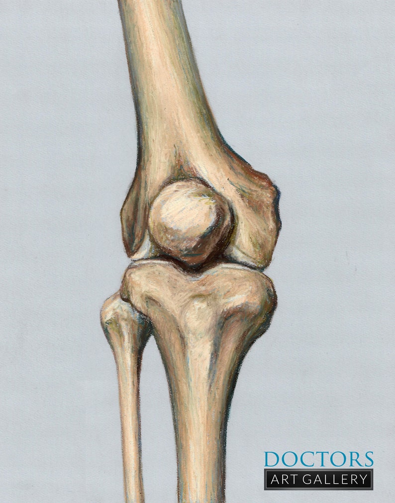
Knee joint Leg Patella Osteology Bone Anatomy Art Print Etsy
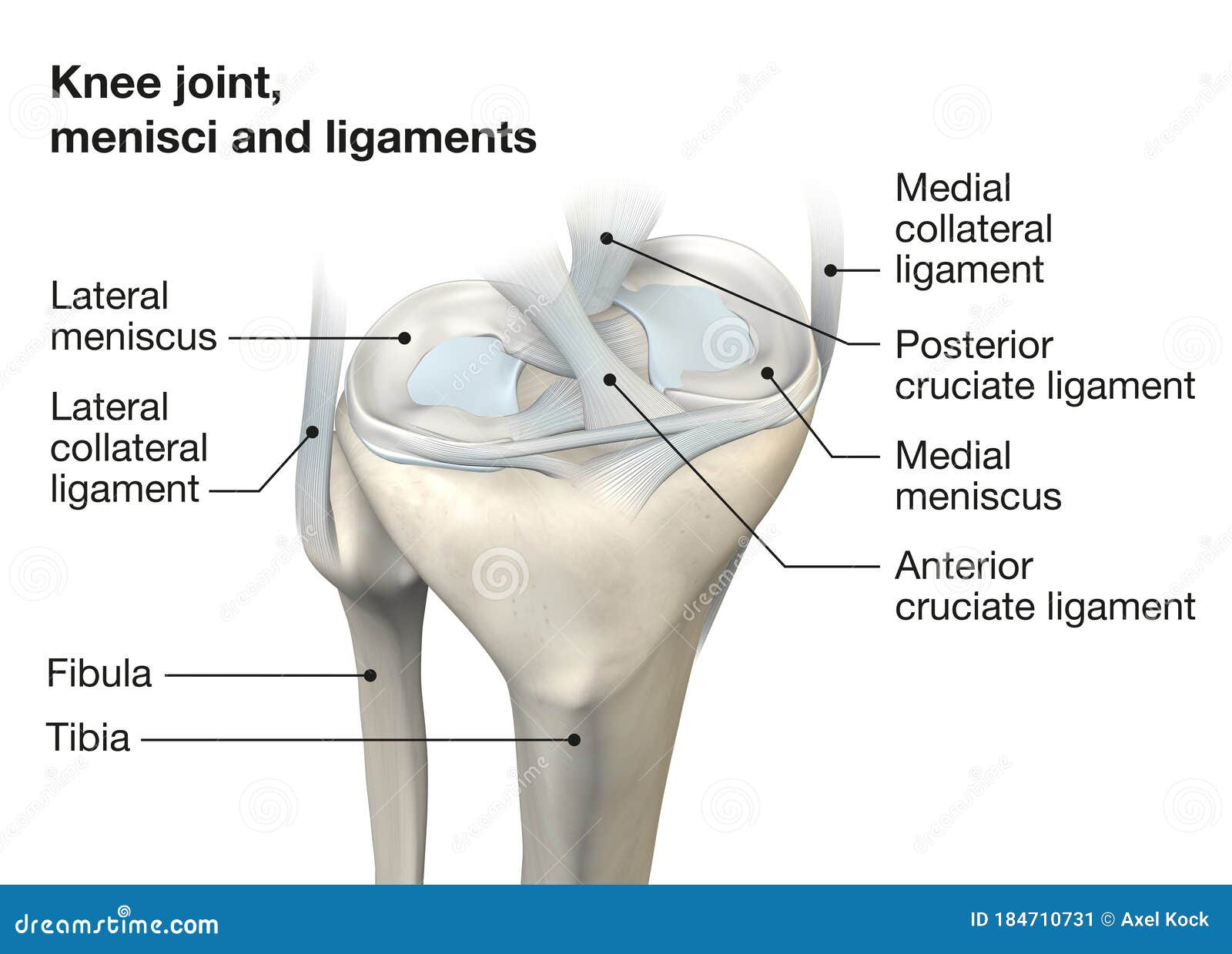
Knee Joint Anatomy, Menisci and Ligaments, Medically 3D Illustration

The Knee Joint Anatomy Sketch

HUMAN KNEE JOINT WITH MAIN BONES. Download Scientific Diagram

Schematic illustration of the knee joint anatomy. Download
Knee Joint from Lateral Surface ClipArt ETC

Ligaments The major ligaments in the knee joint are Patellar ligament
Web Anatomy Of Human Knee Vector Sketch Of Leg Bones And Joint, Medicine Design.
Web Anatomy Of The Knee.
Four Quadriceps Muscles Are Present In Front Of The Knee Which Help In Straightening The Leg From The Knee.
To Understand The Function And Structure Of The Knee Joint, A Knee Anatomy Can Be Helpful.
Related Post: