Pineal Gland Drawing
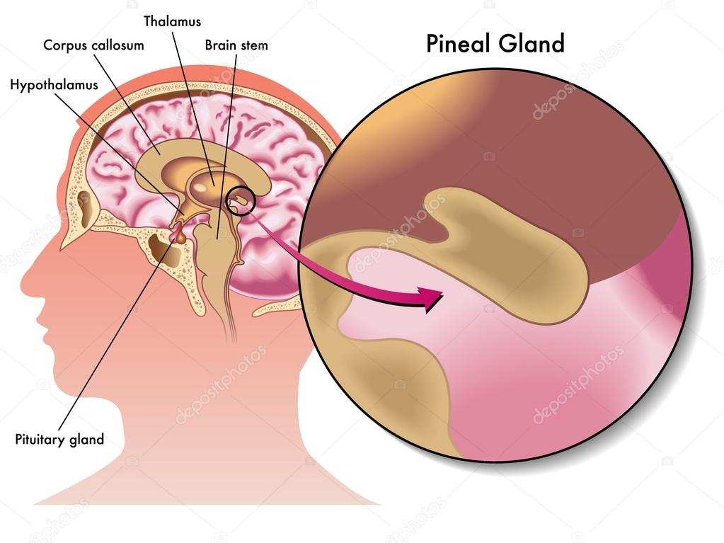
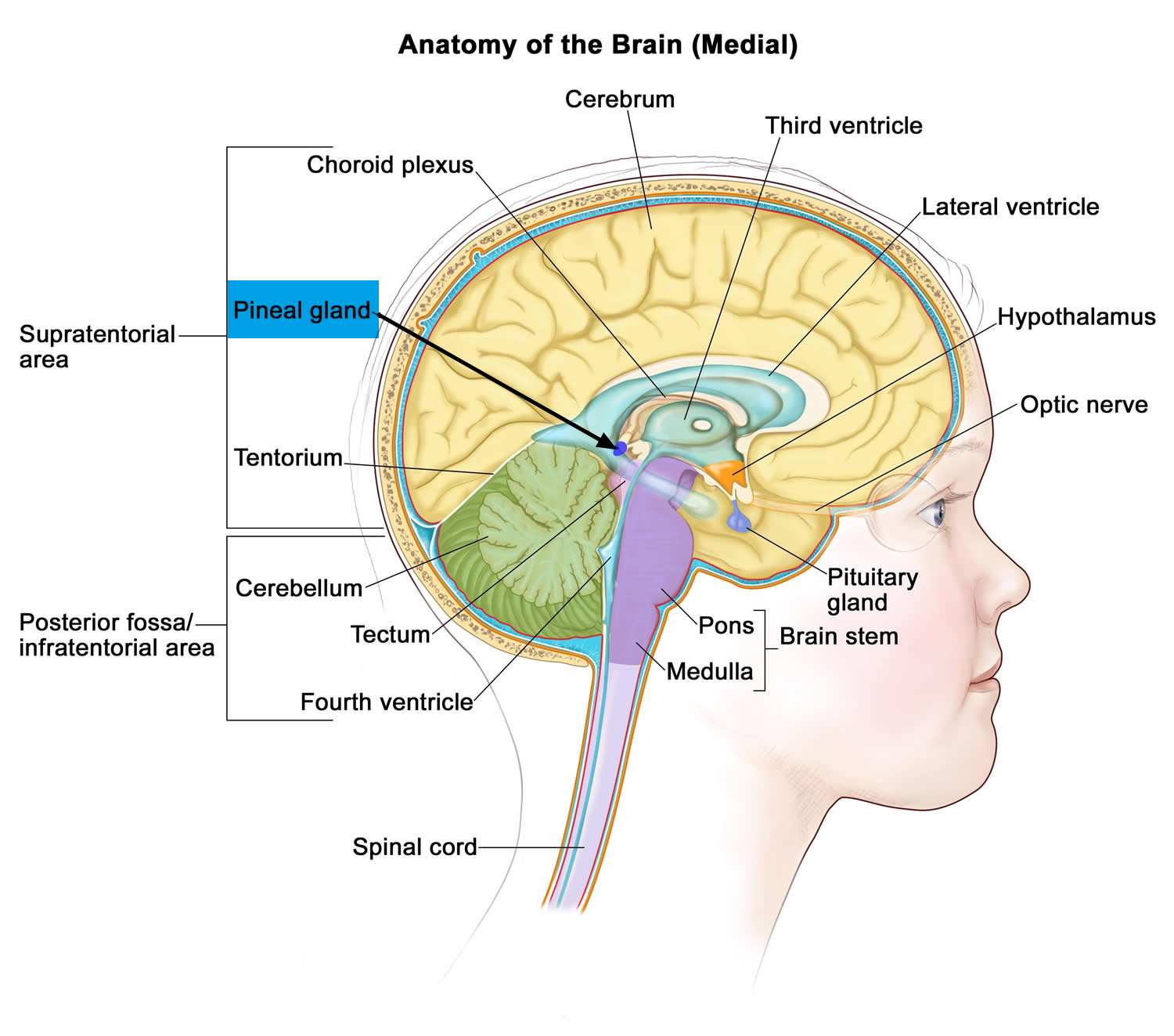
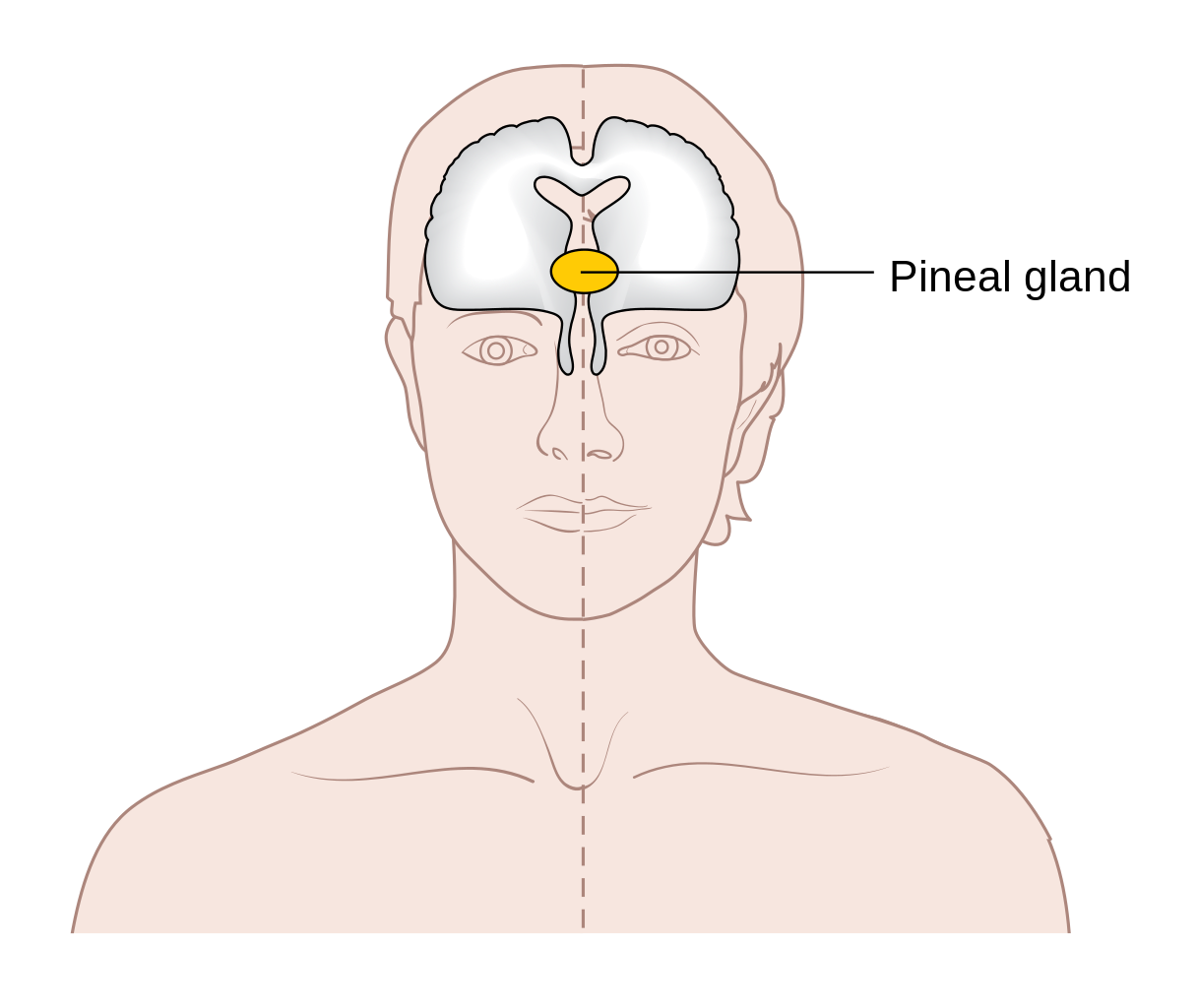
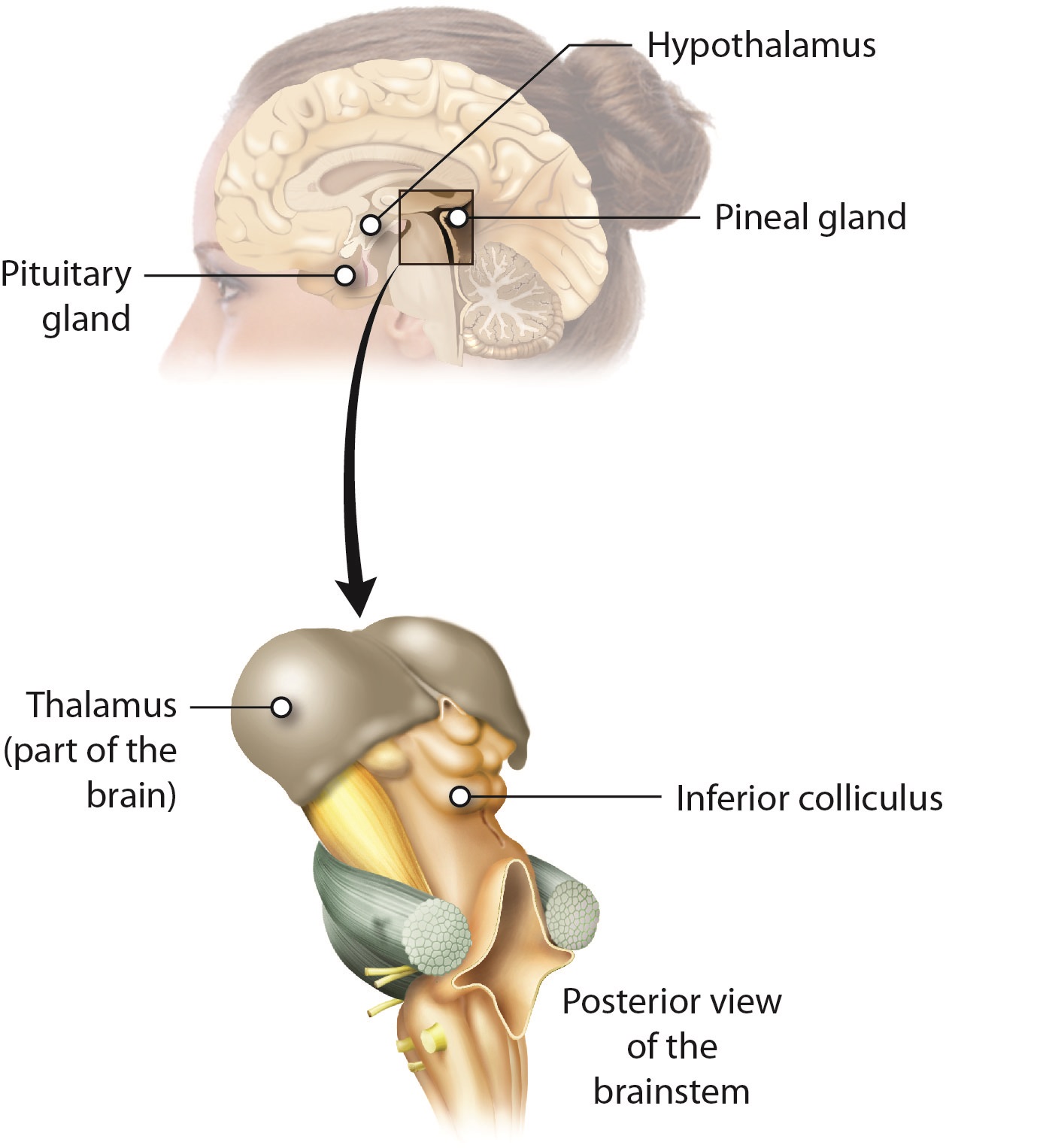
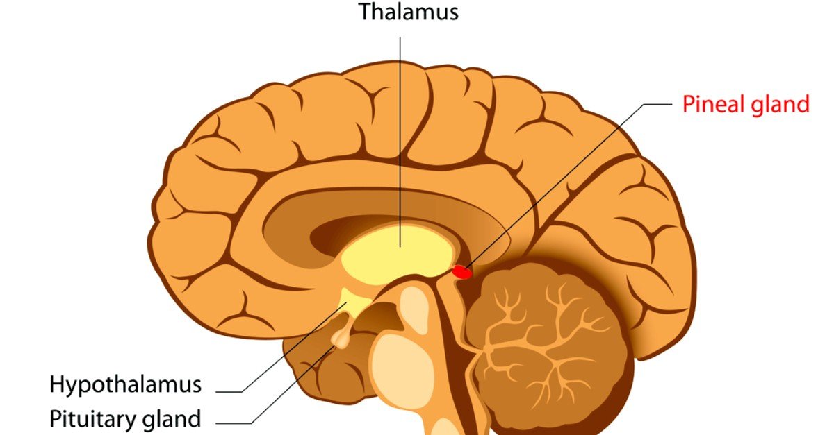
Pineal Gland Drawing - Web the pineal gland is a pine cone shaped structure located in the diencephalon whose main function is the secretion of melatonin, a hormone that is best known. In an adult, it weighs about 0.1 g. Its importance is in the circadian cycle of sleep and wakefulness. Web glands and organs of the endocrine system; The pineal gland, conarium, or epiphysis cerebri. Web the pineal gland develops from the roof of the diencephalon, a section of the brain, and is located behind the third cerebral ventricle in the brain midline (between the two cerebral hemispheres). Histology of pineal gland explanation with step by step drawing, lecture on pineal gland | anatomy | practical | journal drawing | show. 2.6k views 2 years ago endocrine gland histology. The pineal gland, also known as the ‘pineal body,’ is a small endocrine gland. Drawing shows the hypothalamus, pituitary gland, pineal gland, thyroid gland, thymus, adrenal gland, pancreas, ovaries (female), and testes (male). It is also thought to produce hormones that inhibit the action of. There are more than 99,000 vectors, stock photos & psd files. Diagram of pituitary and pineal glands in the human brain. Histology of pineal gland explanation with step by step drawing, lecture on pineal gland | anatomy | practical | journal drawing | show. In humans, this is. Web the pineal gland is a pine cone shaped structure located in the diencephalon whose main function is the secretion of melatonin, a hormone that is best known. Its main secretion is melatonin , which regulates the circadian rhythm of the body. Web pineal gland drawing stock photos and images (141) see pineal gland drawing stock video clips quick filters:. Web [ 1] the anatomy of the pineal gland, along with the pituitary gland, is displayed in the image below. Web sagittal drawing of the brain illustrating the pineal gland () from bock’s 19th century handbuch der anatomie des menschen (1841) leipzig, germany. Web introduction the pineal gland is an endocrine gland located in the posterior aspect of the cranial. Its importance is in the circadian cycle of sleep and wakefulness. 2.6k views 2 years ago endocrine gland histology. The pineal gland, also known as the ‘pineal body,’ is a small endocrine gland. It is also thought to produce hormones that inhibit the action of. Web the pineal gland is located deep in the brain in an area called the. There are more than 99,000 vectors, stock photos & psd files. It is also thought to produce hormones that inhibit the action of. Web the pineal region consists of the two habenular trigones, habenular commissure, pineal body, posterior commissure, and the superior and inferior laminae of the epiphyseal stalk. The pineal gland receives information about light levels in the environment. Drawing shows the hypothalamus, pituitary gland, pineal gland, thyroid gland, thymus, adrenal gland, pancreas, ovaries (female), and testes (male). The pineal gland is also known as the epiphysis cerebri. In humans, this is situated in the m. Autopsy studies have shown that the average size of the pineal gland is similar to that of a grain of rice. In an. It sits in the groove between the two superior colliculi, and is bilaterally related to the posterior aspects of the two thalami. The pineal gland, also known as the ‘pineal body,’ is a small endocrine gland. The pineal gland receives information about light levels in the environment from the. Autopsy studies have shown that the average size of the pineal. There are more than 99,000 vectors, stock photos & psd files. Its importance is in the circadian cycle of sleep and wakefulness. It is also thought to produce hormones that inhibit the action of. The pineal gland, also known as the ‘pineal body,’ is a small endocrine gland. 18 the pineal gland is a small endocrine gland located within the. Web the pineal gland (also known as the pineal body, [1] conarium, or epiphysis cerebri) is a small endocrine gland in the brain of most vertebrates. Web the pineal gland or pineal body is a small gland in the middle of the head. Diagram of pituitary and pineal glands in the human brain. Web glands and organs of the endocrine. The pineal gland is composed of cells called pinealocytes and cells of the nervous system called. In humans, this is situated in the m. Web the pineal gland is located deep in the brain in an area called the epithalamus, where the two halves of the brain join. 2.6k views 2 years ago endocrine gland histology. Web the pineal gland. Its name is derived from its shape, which is similar to that of a pinecone (latin pinea ). The pineal gland is composed of cells called pinealocytes and cells of the nervous system called. Web the pineal region consists of the two habenular trigones, habenular commissure, pineal body, posterior commissure, and the superior and inferior laminae of the epiphyseal stalk. Web the pineal gland is located deep in the brain in an area called the epithalamus, where the two halves of the brain join. The pineal gland, conarium, or epiphysis cerebri. Web you can find & download the most popular pineal gland vectors on freepik. Web the pineal gland develops from the roof of the diencephalon, a section of the brain, and is located behind the third cerebral ventricle in the brain midline (between the two cerebral hemispheres). Autopsy studies have shown that the average size of the pineal gland is similar to that of a grain of rice. Histology of pineal gland explanation with step by step drawing, lecture on pineal gland | anatomy | practical | journal drawing | show. Web [ 1] the anatomy of the pineal gland, along with the pituitary gland, is displayed in the image below. In an adult, it weighs about 0.1 g. Web the pineal gland, also called the pineal body, develops as an outward projection from the posterior wall of the third ventricle, below the splenium of corpus callosum. Drawing shows the hypothalamus, pituitary gland, pineal gland, thyroid gland, thymus, adrenal gland, pancreas, ovaries (female), and testes (male). Web sagittal drawing of the brain illustrating the pineal gland () from bock’s 19th century handbuch der anatomie des menschen (1841) leipzig, germany. Web the pineal gland (also known as the pineal body, [1] conarium, or epiphysis cerebri) is a small endocrine gland in the brain of most vertebrates. There are more than 99,000 vectors, stock photos & psd files.
Know your brain Pineal gland — Neuroscientifically Challenged
Pineal gland — Stock Vector © rob3000 65937043

PINEAL GLAND, DRAWING Stock Photo Alamy
![]()
Pineal gland Anatomy, histology and blood supply Kenhub

Pineal gland, illustration Stock Image F036/1616 Science Photo
Pineal Gland & its Function Cyst & Calcified Pineal Gland
What Part Of The Brain Controls The Pineal Gland
KnowledgeWorks Drawing Pineal gland and thalamus English labels
The Meaning Of The Pineal Gland Spirit Molecule

Anatomy of pathway of stimulation of the pineal gland. CN, conarii or
The Pineal Gland Is Also Known As The Epiphysis Cerebri.
It Is Located On The Back Portion Of The Third Cerebral Ventricle Of The Brain, Which Is A Fluid.
Web The Pineal Gland Is A Small Neuroendocrine Organ In The Diencephalon Region Of The Brain.
The Pineal Gland Receives Information About Light Levels In The Environment From The.
Related Post: