Retina Drawing Template
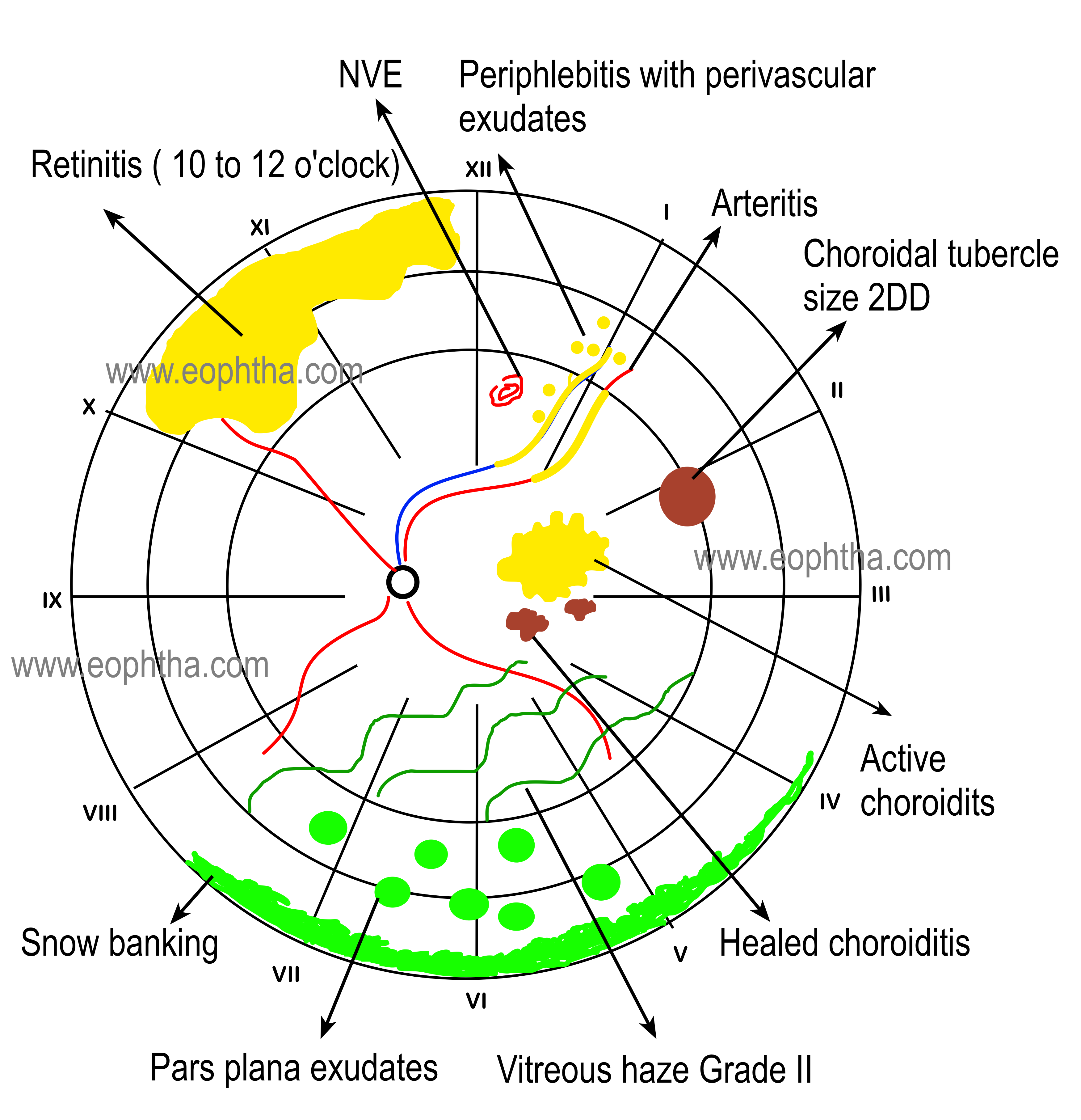
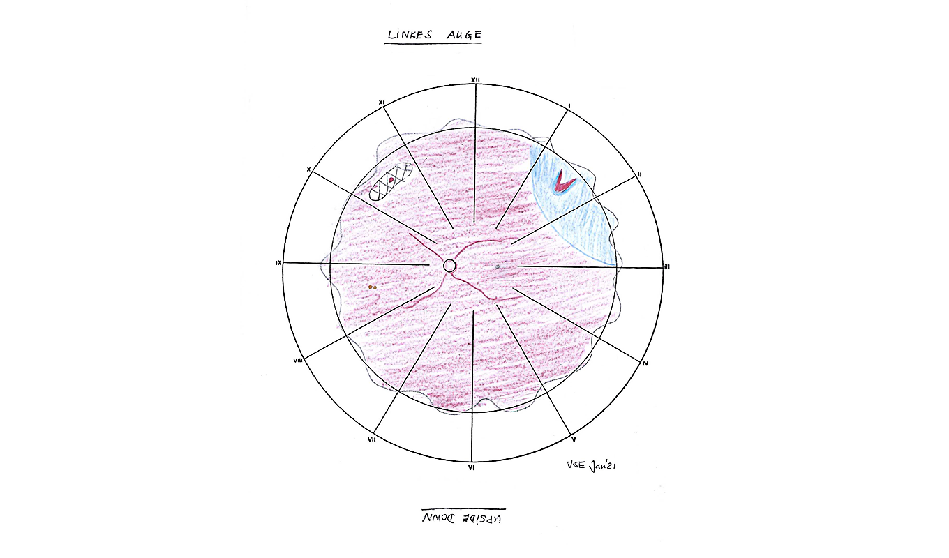
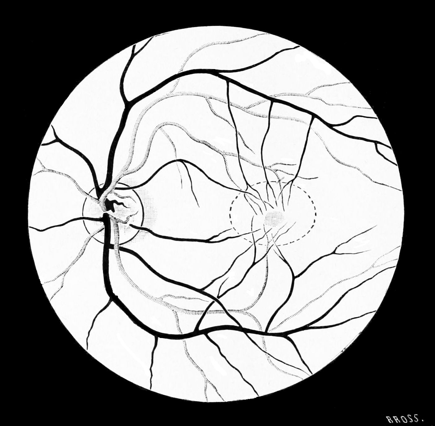
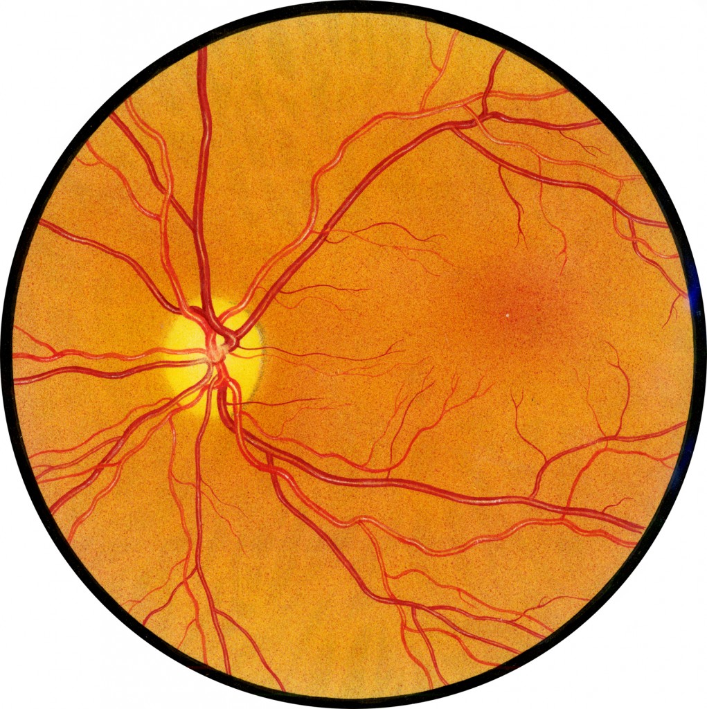
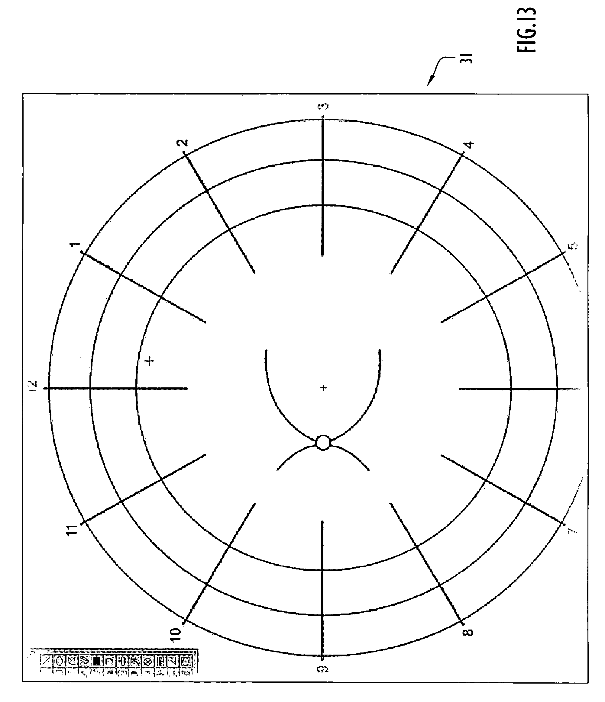
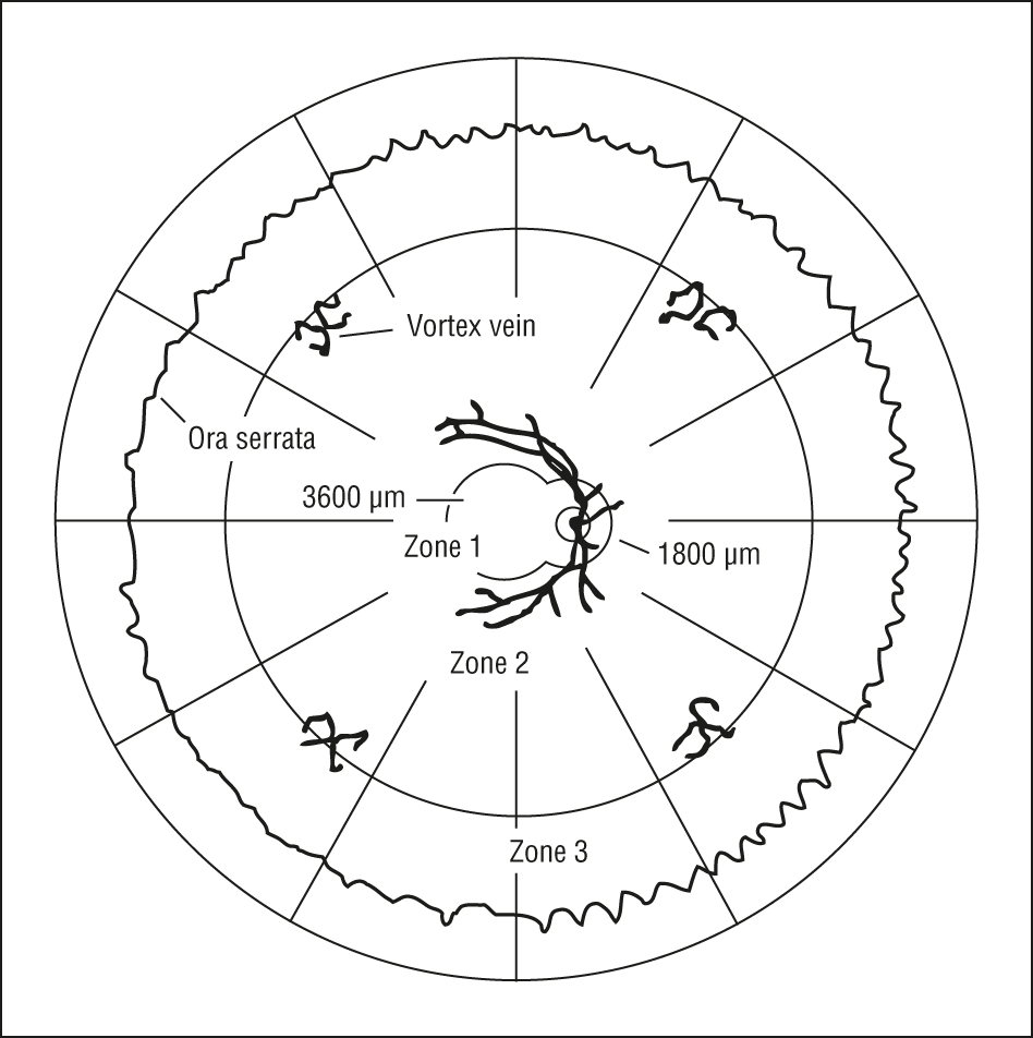
Retina Drawing Template - Advantages of indirect ophthalmoscopy, optical principle, fundus drawing. Below you’ll see thumbnail sized previews of the title slides of a few of our 68 best retina templates for powerpoint and google slides. Web requisite for drawing: Indirect ophthalmoscopy and fundus drawing. Draw it on the right for the right eye, and on the left for the left eye. The iris is the colored part of the eye that regulates the amount of light entering the eye. Web pdf | on jul 1, 2011, luann dvorak and others published retinal drawing: The best free retinal drawing images. Documentation must be legible retinal drawing must be maintained in the patient’s record drawings should include sufficient detail, standard colors, and appropriate labels individual for each eye Web here presented 39+ retinal drawing images for free to download, print or share. Next, draw the macula temporal to it. (see a great example here ). Retinal drawing | free download on clipartmag. Retinal drawings deviated significantly from the ideal no difference between drawing and objectively measured rrd border distance from the fovea (p < 0.0001). Web here presented 39+ retinal drawing images for free to download, print or share. Although payer policies differ, some common charting requirements include the following: A true retina drawing will contain three concentric circles. It is located in the center of the retina. Learn how to draw retinal pictures using these outlines or print just for coloring. Web here presented 39+ retinal drawing images for free to download, print or share. Web a detailed retinal drawing is required. It’s also a great activity for you to do to help you relax and develop a new skill. Next, draw the macula temporal to it. Although payer policies differ, some common charting requirements include the following: Web pdf | on jul 1, 2011, luann dvorak and others published retinal drawing: Download from 18 free drawings of. Documentation must be legible retinal drawing must be maintained in the patient’s record drawings should include sufficient detail, standard colors, and appropriate labels individual for each eye Retinal drawings deviated significantly from the ideal no difference between drawing and objectively measured rrd border distance from the fovea (p < 0.0001). You can use a. To allow modifications of drawing some stencils: Although payer policies differ, some common charting requirements include the following: Web start with your pupils and the indirect ophthalmoscope, then add your arms, the patient’s pupil and the indirect lens, thus making your pupil and your depressor a straight line and using the patient’s pupil as a fulcrum. Download from 18 free. Advantages of indirect ophthalmoscopy, optical principle, fundus drawing. Web the fundus skeleton. Web here presented 39+ retinal drawing images for free to download, print or share. Retinal drawings deviated significantly from the ideal no difference between drawing and objectively measured rrd border distance from the fovea (p < 0.0001). Web looking deep into retina : Web looking deep into retina : Ehr perils and recommendations peril: You can use a circular object cornea & anterior segment: Web the requisites for drawing include six colored pencils or pens such as black, blue, brown, red, green, and yellow and an eraser to allow modifications of drawing. Indirect ophthalmoscopy and fundus drawing. Below you’ll see thumbnail sized previews of the title slides of a few of our 68 best retina templates for powerpoint and google slides. Although payer policies differ, some common charting requirements include the following: Overview of main colors retinado d 217 subscribers subscribe 34 views 1 year ago lets take a look into the most important colors for retinal. There are some stencils available commercially which aid in the quick reproduction of cornea and retina diagrams, front of eye, and slit shape outlines. The best free retinal drawing images. Documentation must be legible retinal drawing must be maintained in the patient’s record drawings should include sufficient detail, standard colors, and appropriate labels individual for each eye You can use. Advantages of indirect ophthalmoscopy, optical principle, fundus drawing. Web the fundus skeleton. To allow modifications of drawing some stencils: Web here presented 39+ retinal drawing images for free to download, print or share. (see a great example here ). Web a detailed retinal drawing is required. Retinal drawings deviated significantly from the ideal no difference between drawing and objectively measured rrd border distance from the fovea (p < 0.0001). The iris is the colored part of the eye that regulates the amount of light entering the eye. Ehr perils and recommendations peril: Web documentation of medical necessity; The best free retinal drawing images. Retinal drawing | free download on clipartmag. You can use a circular object cornea & anterior segment: Although payer policies differ, some common charting requirements include the following: Indirect ophthalmoscopy and fundus drawing. Web an extensive scaled drawing must accurately represent normal, abnormal, and common findings such as lattice degeneration, hypertensive vascular changes, proliferative diabetic retinopathy, as well as retinal detachment, holes, tears, or tumors. Horseshoe shapes at 1:30 with accompanying ablatio retinae from 1 to 3 o’clock and degeneration area at 10:30 with a small round hole first, trace the optic nerve on the retinal drawing. There are some stencils available commercially which aid in the quick reproduction of cornea and retina diagrams, front of eye, and slit shape outlines. Web the requisites for drawing include six colored pencils or pens such as black, blue, brown, red, green, and yellow and an eraser to allow modifications of drawing. Web the fundus skeleton. A drawing that is clearly identified, labeled, and appropriately represents the retinal pathology;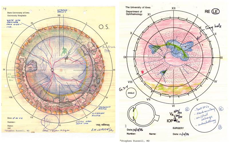
Retinal Drawing at GetDrawings Free download
Retinal Drawing at GetDrawings Free download
Retinal Drawings HEINE Optotechnik
Retinal Drawing at GetDrawings Free download

Documentation & Drawing in Ophthalmology

My efforts as an artist learning to draw the retina — Matt Weed, MD
Retinal Drawing Free download on ClipArtMag
Retinal Drawing at Explore collection of Retinal
The best free Retinal drawing images. Download from 18 free drawings of
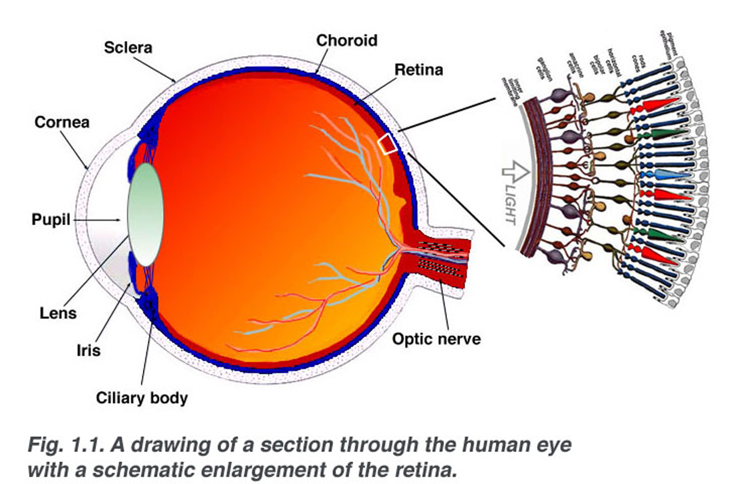
Simple Anatomy of the Retina by Helga Kolb Webvision
To Allow Modifications Of Drawing Some Stencils:
Download From 18 Free Drawings Of.
Eye Drawings Look Artistic And Often Look Complicated, But They’re Actually Quite Easy To Do.
Draw It On The Right For The Right Eye, And On The Left For The Left Eye.
Related Post: