Skin Cell Drawing
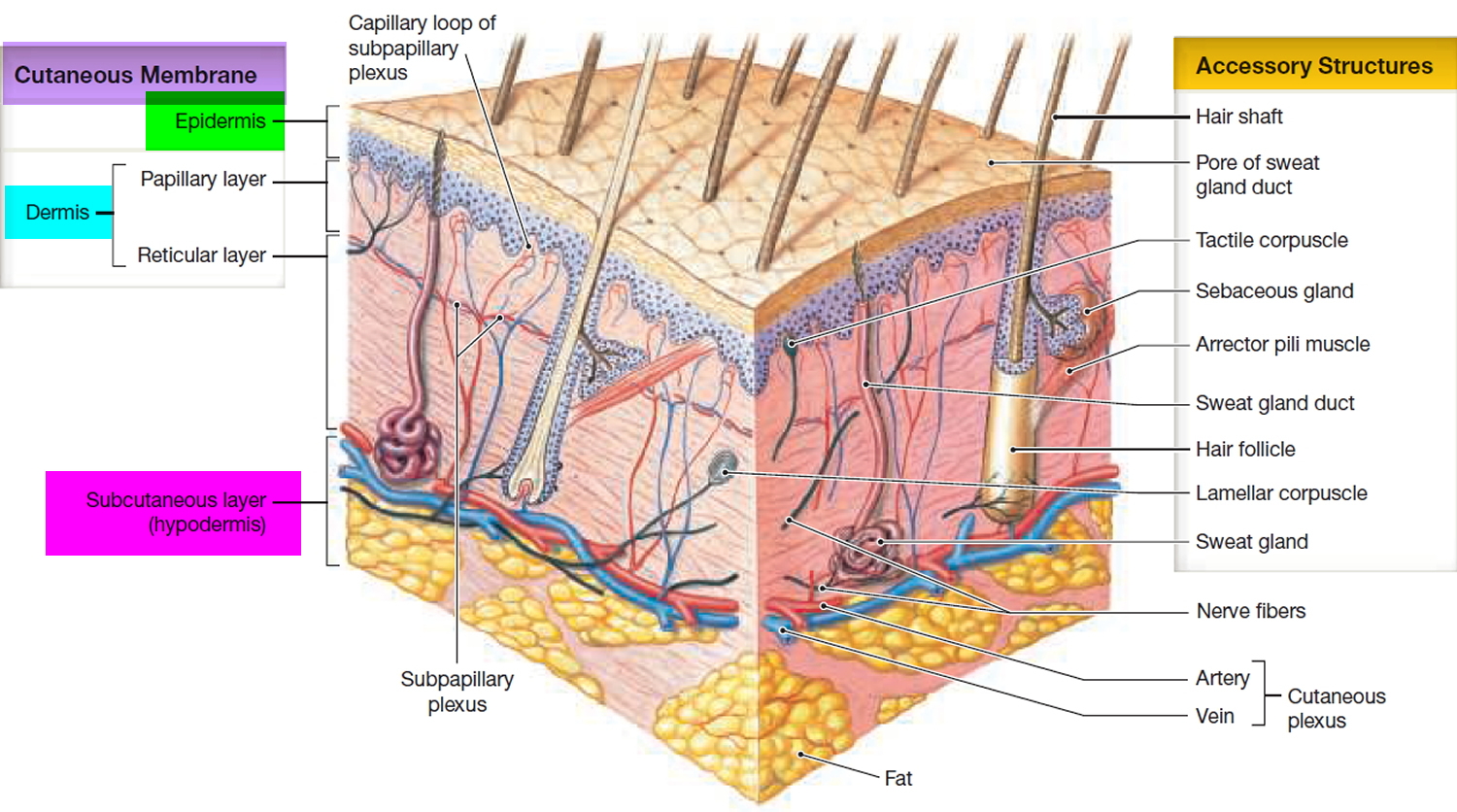
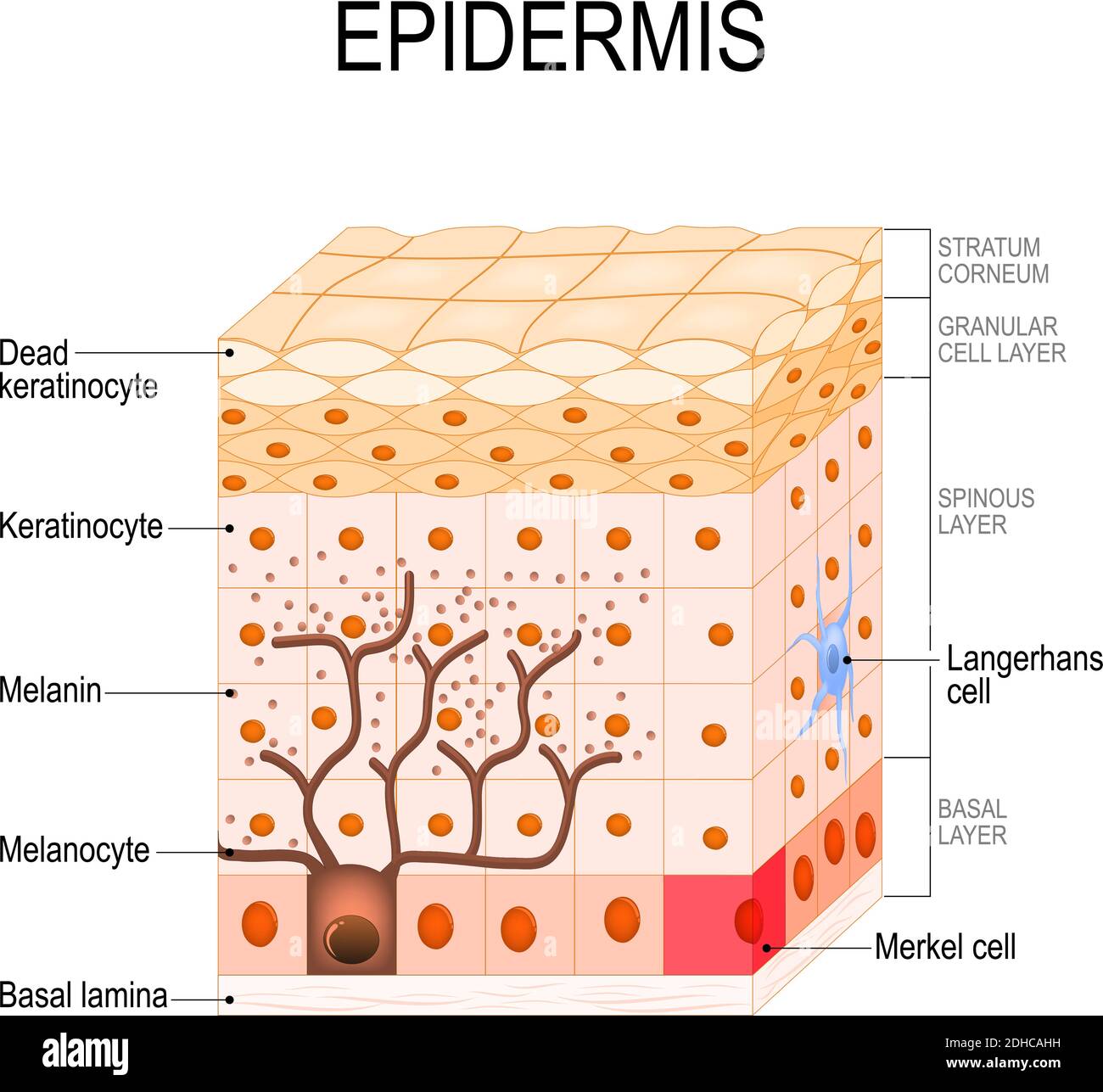
Skin Cell Drawing - Internal organs of the human body. Web the epidermis the dermis hypodermis the number of skin layers that exists depends on how you count them. Web skin that has four layers of cells is referred to as “thin skin.” from deep to superficial, these layers are the stratum basale, stratum spinosum, stratum granulosum, and stratum corneum. Nerves in the skin help you feel sensations like hot and cold. The outermost layer is continuously shed is called the stratum corneum. This gives the skin its color. Human skin cells healthy skin cells skin cells microscope dead skin cells skin cells vector 3d skin cells skin cells close up skin cells background Basal cells are found just under the squamous cells, at the base of the epidermis. Web (usmle topics) structure of the skin, layers of the epidermis, skin barrier and pigmentation. Web choose from drawing of a skin cells stock illustrations from istock. Web (usmle topics) structure of the skin, layers of the epidermis, skin barrier and pigmentation. Web the deepest layer of the epidermis is the stratum basale (stratum germinativum), a layer of stem cells that produce all of the keratinocytes, or skin cells, in the epidermis. Each type of skin cell has a unique role that contributes to the overall structure. Layers of epidermis illustration vector on white background. Your skin protects your body from germs and regulates body temperature. Vector illustration of young and aged skin. Keratin strengthens the skin and makes it waterproof. Each type of skin cell has a unique role that contributes to the overall structure and function of. The epidermis, made of closely packed epithelial cells, and the dermis, made of dense, irregular connective tissue that houses blood vessels, hair follicles, sweat glands, and other structures. Web overview the three layers of skin on top of muscle tissue. Web diagram of human skin structure. Human gas exchange process with oxygen cycle explanation outline diagram. Web this article describes. The skin is the body’s largest organ, made of water, protein, fats and minerals. Web (usmle topics) structure of the skin, layers of the epidermis, skin barrier and pigmentation. Web diagram of human skin structure. The epidermis, made of closely packed epithelial cells, and the dermis, made of dense, irregular connective tissue that houses blood vessels, hair follicles, sweat glands,. Melanocytes are found at the base of the epidermis and make melanin. Web skin that has four layers of cells is referred to as “thin skin.” from deep to superficial, these layers are the stratum basale, stratum spinosum, stratum granulosum, and stratum corneum. Layers of epidermis illustration vector on white background. Melanocytes that produce melanin are also present in this. The cells in this layer are called keratinocytes. Skin cell diagram stock photos are available in a variety of sizes and formats to fit your needs. Internal organs of the human body. Within these layers are additional layers. It consists of 2 primary types of cells: Web find & download the most popular skin cell vectors on freepik free for commercial use high quality images made for creative projects. Skin cell diagram stock photos are available in a variety of sizes and formats to fit your needs. Most popular sectional view of the skin.comparison illustration of protection. The keratinocytes are composed of a protein called keratin.. Web choose from drawing of a skin cells stock illustrations from istock. The epidermis, made of closely packed epithelial cells, and the dermis, made of dense, irregular connective tissue that houses blood vessels, hair follicles, sweat glands, and other structures. Web the epidermis is the thin outer layer of the skin. Melanocytes that produce melanin are also present in this. Melanocytes are also found at the base of the epidermis and make melanin. If you count the layers within the layers, the skin has eight or even 10 layers. The epidermis is a tough coating formed from overlapping layers of dead skin cells. Web hypodermis epidermis it is the outermost layer of the skin. You have three main layers of. Nerves in the skin help you feel sensations like hot and cold. These are keratinocytes, melanocytes, langerhans cells, and merkel cells. If you count the layers within the layers, the skin has eight or even 10 layers. Web the epidermis is the thin outer layer of the skin. Web this tutorial shows scientists how to draw skin cells in adobe. Web choose from drawing of a skin cells stock illustrations from istock. Melanocytes are found at the base of the epidermis and make melanin. Human gas exchange process with oxygen cycle explanation outline diagram. Web the epidermis the dermis hypodermis the number of skin layers that exists depends on how you count them. Purchase pdf (script of this video + images) here: “thick skin” is found only on the palms of the hands and the soles of the feet. You have three main layers of skin—the epidermis , dermis, and hypodermis (subcutaneous tissue). Within these layers are additional layers. Web (usmle topics) structure of the skin, layers of the epidermis, skin barrier and pigmentation. This gives the skin its color. Layers of epidermis illustration vector on white background. Web this article describes the histology of the skin, including layers, cell types, contents and characteristics. Each type of skin cell has a unique role that contributes to the overall structure and function of. The skin is the body’s largest organ, made of water, protein, fats and minerals. Web the term ‘skin cell’ may refer to any of the four main types of cells found in the epidermis. These are keratinocytes, melanocytes, langerhans cells, and merkel cells.
epidermal skin cells About Dermatology
Schematic representation of skin structure and cell population. The

human skin cells labeled Google Search Acanthosis Nigricans, Skin
Epidermis 5 Layers of Epidermis, Outermost Layer & Function

how to draw skin cell diagram YouTube

Layers of epidermis vector illustration Skin anatomy, Epidermis, Skin

Skin Structure infographic LifeMap Discovery
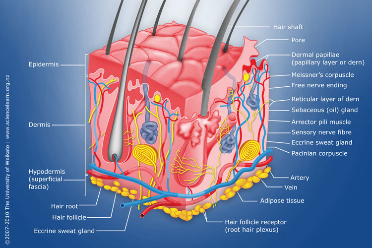
Diagram of human skin structure — Science Learning Hub
Skin cells microscope hires stock photography and images Alamy
The Structure Of Human Skin Cells Stock Illustration Download Image
Nerves In The Skin Help You Feel Sensations Like Hot And Cold.
Skin Cell Diagram Stock Photos Are Available In A Variety Of Sizes And Formats To Fit Your Needs.
The Epidermis Is A Tough Coating Formed From Overlapping Layers Of Dead Skin Cells.
Diagram Of Human Cell For Education Illustration.
Related Post:
