Skull Anterior View Drawing
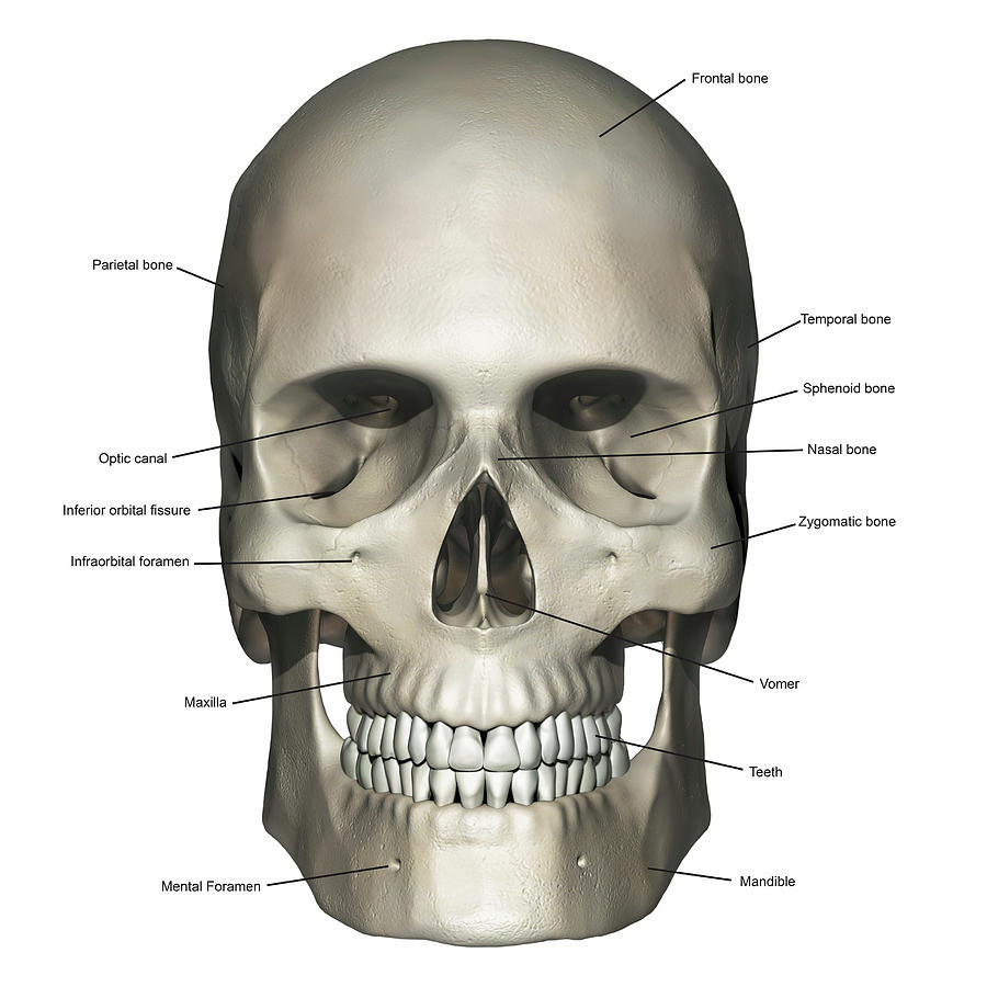
Skull Anterior View Drawing - An anterior view of the skull shows the bones that form the forehead, orbits (eye sockets), nasal cavity, nasal septum, and upper and lower jaws. The anterior and posterior spinal arteries also. This is a model of the human (homo sapiens) skull. Smartdraw includes 1000s of professional healthcare and anatomy chart templates that you can modify and make your own. Inside the nasal area of the skull, the nasal cavity. Web anterior view of skull. The major anterior skull bones include: Just posterior to the frontal. The idea behind using labeled diagrams is to get an overview of all of the structures within a given area. And we continue to move forward with thes. The outside of the cranial base is called the external cranial base. This drawing of the skull shows the bones that form the cranium and mandible. Overview image of an anterior view of the skull. Web the bones of the skull that are visible from an anterior and a lateral view are the following: And we continue to move forward. Smartdraw includes 1000s of professional healthcare and anatomy chart templates that you can modify and make your own. The sphenoid bone (with the greater and the lesser wings) the frontal bone (especially the orbital surface) the zygomatic bone; Inside the nasal area of the skull, the nasal cavity is divided into halves by the nasal septum. This is a model. This image by the royal college of surgeons of ireland (rcsi) is retrieved from health education assets library (heal) of the university of utah. This is a model of the human (homo sapiens) skull. Inside the nasal area of the skull, the nasal cavity is divided into halves by the nasal septum. Web the bones of the skull that are. Inside the nasal area of the skull, the nasal. Web the bones of the skull that are visible from an anterior and a lateral view are the following: Web anterior to this we have the superior surface of the orbital plate, which is the region on which the frontal lobe rests. An anterior view of the skull shows the bones. The anterior and posterior spinal arteries also. This is a tutorial about human anatomy. Inside the nasal area of the skull, the nasal cavity. This drawing of the skull shows the bones that form the cranium and mandible. Web labeled skull diagram. It allows the spinal cord to pass inferiorly out of the cranial vault, and also the vertebral arteries to enter the skull and provide the posterior input to the circle of willis. Inside the nasal area of the skull, the nasal cavity. The major anterior skull bones include: The cranial bones surround and protect the brain and house the middle. This is a model of the human (homo sapiens) skull. This drawing of the skull shows the bones that form the cranium and mandible. Web figure 7.4 anterior view of skull an anterior view of the skull shows the bones that form the forehead, orbits (eye sockets), nasal cavity, nasal septum, and upper and lower jaws. Inside the nasal area. Web human head (anterior view) the human head is more than just a nuisance responsible for your headaches. Just posterior to the frontal. Inside the nasal area of the skull, the nasal. The cranial bones surround and protect the brain and house the middle and inner ear structures. It allows the spinal cord to pass inferiorly out of the cranial. The outside of the cranial base is called the external cranial base. Web it is subdivided into the facial bones and the cranial bones. Smartdraw includes 1000s of professional healthcare and anatomy chart templates that you can modify and make your own. How to draw the skull from front and side so easy and uncomplicated. Web the skull (cranium) is. The anterior and posterior spinal arteries also. How to draw the skull from front and side so easy and uncomplicated. In the ct data, the upper teeth and lower teeth were joined as one mesh, therefore i completely remodelled some of the teeth so that the mandible could be. The facial bones underlie the facial structures, form the nasal cavity,. Frontal bone, mandible, maxilla, nasal bone, parietal bone, temporal bone, and zygomatic bone. The idea behind using labeled diagrams is to get an overview of all of the structures within a given area. The sphenoid bone (with the greater and the lesser wings) the frontal bone (especially the orbital surface) the zygomatic bone; Then add lines that show where the jawbones come down to. Inside the nasal area of the skull, the nasal. Web osteology of the skull objectives • to name the bones of the neurocranium and viscerocranium • to identify the various bones and sutures as seen from anterior, lateral, posterior, inferior, and interior views of the skull • to name the openings, foramina, and canals as seen from the aforementioned views The major anterior skull bones include: This is a tutorial about human anatomy. Web figure 7.4 anterior view of skull an anterior view of the skull shows the bones that form the forehead, orbits (eye sockets), nasal cavity, nasal septum, and upper and lower jaws. The anterior and posterior spinal arteries also. Draw a wide oval to represent the top portion of the skull. The foramen of the anterior skull include (top to bottom): From openstax book 'anatomy and physiology', fig. An anterior view of the skull shows the bones that form the forehead, orbits (eye sockets), nasal cavity, nasal septum, and upper and lower jaws. The cranial bones surround and protect the brain and house the middle and inner ear structures. It was then cleaned, adapted and polypainted in zbrush.
The Bones of the Skull Human Anatomy and Physiology Lab (BSB 141)

The Skull Anatomy and Physiology I

Anterior view Skull Netter Skull anatomy, Craniosacral therapy
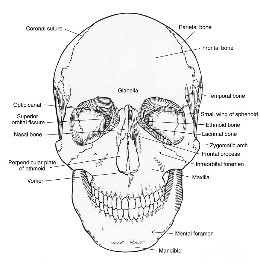
Illustration Of Anterior Skull Photograph by Science Source Pixels

1. Selected anatomical landmarks of the skull (anterior view

Printable Nursing and NCLEX
Anterior View Of Human Skull Anatomy Photograph by Alayna Guza Fine

Anatomy of Skull Illustration Anterior View Labelled Medical Stock
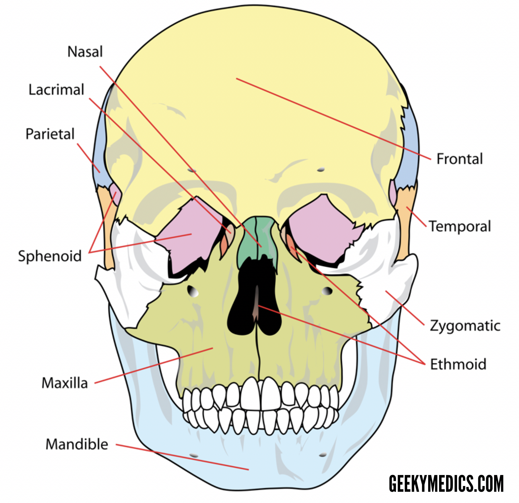
Bones of the Skull Skull Osteology Anatomy Geeky Medics
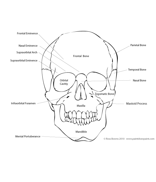
Paint Draw Paint, with Ross Bowns Drawing Basics Anatomy of the Skull
In The Ct Data, The Upper Teeth And Lower Teeth Were Joined As One Mesh, Therefore I Completely Remodelled Some Of The Teeth So That The Mandible Could Be.
In The Anterior Most Part Of The Anterior Cranial Fossa In The Midline, Is A Small Triangular Bump Known As The Frontal Crest, And It Is Structurally Continuous With The Superior Sagittal Sinus.
How To Draw The Skull From Front And Side So Easy And Uncomplicated.
Web An Anterior View Of The Skull Shows The Bones That Form The Forehead, Orbits (Eye Sockets), Nasal Cavity, Nasal Septum, And Upper And Lower Jaws.
Related Post: